. 2021 Dec 22;6(24):e150451.
doi: 10.1172/jci.insight.150451.

Hypoxia induces DOT1L in articular cartilage to protect against osteoarthritis

Affiliations

Hypoxia induces DOT1L in articular cartilage to protect against osteoarthritis

Astrid De Roover et al. JCI Insight. .

Abstract

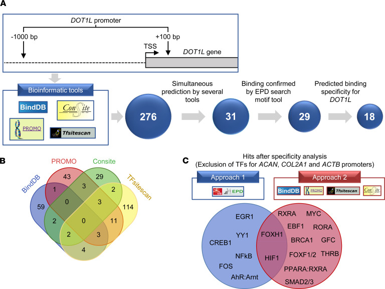
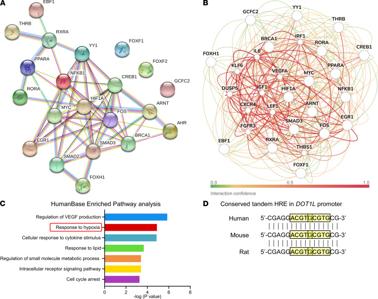
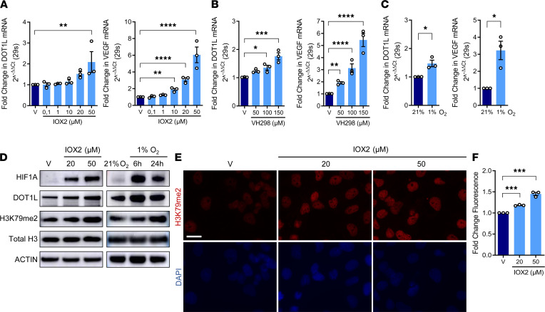
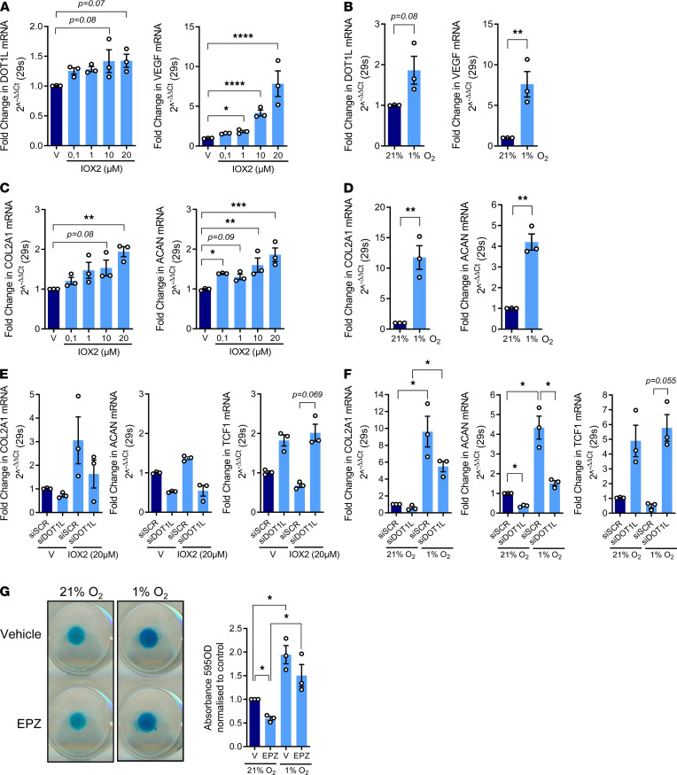
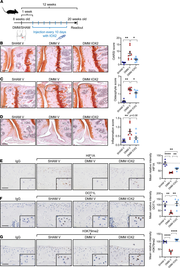
Osteoarthritis is the most prevalent joint disease worldwide, and it is a leading source of pain and disability. To date, this disease lacks curative treatment, as underlying molecular mechanisms remain largely unknown. The histone methyltransferase DOT1L protects against osteoarthritis, and DOT1L-mediated H3K79 methylation is reduced in human and mouse osteoarthritic joints. Thus, restoring DOT1L function seems to be critical to preserve joint health. However, DOT1L-regulating molecules and networks remain elusive, in the joint and beyond. Here, we identified transcription factors and networks that regulate DOT1L gene expression using a potentially novel bioinformatics pipeline. Thereby, we unraveled a possibly undiscovered link between the hypoxia pathway and DOT1L. We provide evidence that hypoxia enhanced DOT1L expression and H3K79 methylation via hypoxia-inducible factor-1 α (HIF1A). Importantly, we demonstrate that DOT1L contributed to the protective effects of hypoxia in articular cartilage and osteoarthritis. Intra-articular treatment with a selective hypoxia mimetic in mice after surgical induction of osteoarthritis restored DOT1L function and stalled disease progression. Collectively, our data unravel a molecular mechanism that protects against osteoarthritis with hypoxia inducing DOT1L transcription in cartilage. Local treatment with a selective hypoxia mimetic in the joint restores DOT1L function and could be an attractive therapeutic strategy for osteoarthritis.

Keywords: Aging; Bone Biology; Cartilage; Osteoarthritis.


Figures

Figure 1
Figure 1. Bioinformatics pipeline identifies transcription factors regulating the DOT1L gene.
(A) Overview of the bioinformatics analysis flow of the human DOT1L proximal promoter. The top part of A displays the DOT1L gene promoter region that was used for the analysis, namely –1000 bp to +100 bp relative to the transcription start site (TSS). The bottom part shows the 4 different bioinformatics web-based tools that were used and the transcription factor (TF) selection pipeline. (B) Venn diagram of the 276 TFs found by the 4 different tools, of which the TFs predicted by at least 2 different tools were selected for further analysis. (C) Overview of hits remaining after the specificity analysis. Two different approaches were used to determine whether the TFs were more specific for the DOT1L promoter compared with the aggrecan (ACAN), collagen 2a1 (COL2A1), and actin (ACTB) promoters. The diagram shows the 18 resulting TFs predicted to be more specific for DOT1L after exclusion of TFs by approach 1 or 2 as well as their overlap.
Figure 2
Figure 2. Bioinformatics analysis unravels an undiscovered link between hypoxia and DOT1L.
(A) STRINGdb protein-protein network of the 18 resulting transcription factors (TFs) upon the specificity analysis. (B and C) Cartilage-specific gene network of the 18 resulting TFs upon the specificity analysis using HumanBase (GIANT) (B) and its pathway enrichment analysis (C). (D) Presence of tandem hypoxia-response elements (HREs) with consensus sequence 5′-(A/G)CGTG-3′ (highlighted with yellow boxes) within the human, mouse, and rat DOT1L gene promoters.
Figure 3
Figure 3. Hypoxia increases DOT1L and H3K79 methylation in human articular chondrocytes.
(A and B) Real-time PCR for DOT1L and VEGF in C28/I2 cells treated with IOX2, VH298, or vehicle (V) for 72 hours (C28/I2, *P < 0.05, **P < 0.01, ***P < 0.001, ****P < 0.0001 in A and B, Holm corrected for 16 and 6 tests by generalized least squares model). (C) Real-time PCR in normoxic (21% O2) or hypoxic (1% O2) conditions for 6 hours (n = 3, *P < 0.05 by Welch-corrected t test). (D) Immunoblot of hypoxia-inducible factor-1 α (HIF1A), DOT1L, and H3K79 methylation after IOX2 treatment for 96 hours and in response to hypoxia. Images are representative of 2 independent experiments. (E and F) H3K79 methylation by immunofluorescence (red) and DAPI staining (blue) in C28/I2 cells after 72 hours. Images are representative of 3 independent experiments with technical duplicates. Scale bar: 50 μm. Fluorescence intensity per cell relative to V (n = 20 images per condition for each experiment; n = 3, ***P < 0.001, Holm corrected for 3 tests by generalized least squares model by generalized least squares model).
Figure 4
Figure 4. Hypoxia-mediated induction of DOT1L depends on HIF1A but not HIF2A.
(A) Luciferase assay in C28/I2 cells transfected with empty plasmid, full DOT1L promoter reporter, or negative control shorter DOT1L-promoter reporter, without conserved overlapping tandem HREs, upon treatment with IOX2 (20μM) for 72 hours, normalized to total protein relative to empty plasmid and vehicle (V) (n = 3, *P < 0.05, **P < 0.01, Šidák corrected for 6 tests in 2-way ANOVA). (B) Real-time PCR with siRNA-mediated silencing of HIF1A, HIF2A, or scrambled control (siSCR) (n = 3, *P < 0.05, **P < 0.01, ****P < 0.0001, Šidák corrected for 6 tests in 1-way ANOVA). (C) ChIP quantitative PCR (ChIP-qPCR) for HIF1A and HIF2A binding to DOT1L and VEGF promoters in cells treated with IOX2 (20 μM) for 72 hours (n = 3, *P < 0.05 by 1-sided t test). (D) MACS2-binding scores around DOT1L and VEGF transcription start site (TSS) of publicly available HIF1A and HIF2A ChIP-Seq data (ChIP-Atlas database). (E) Visualization of HIF1A ChIP-Seq in various cells around the DOT1L TSS. Box indicates overlapping HREs. ChIP-Atlas data mapped to reference human genome (hg19) using Integrative Genomics Viewer (IGV). Data are shown as the mean ± SEM.
Figure 5
Figure 5. DOT1L contributes to the protective effects of hypoxia in human articular chondrocytes.
(A) Real-time PCR analysis of DOT1L and VEGF in primary human articular chondrocytes (hACs) after treatment with hypoxia mimetic IOX2 or vehicle (V) at the indicated concentrations for 72 hours (n = 3, *P < 0.05, ****P < 0.0001, Holm corrected for 10 tests by generalized least squares model). (B) Real-time PCR analysis of DOT1L and VEGF in primary hACs cultured in normoxic (21% O2) or hypoxic (1% O2) conditions for 14 days (n = 3, **P < 0.01 by paired t test). (C) Real-time PCR analysis of COL2A1 and ACAN in primary hACs after treatment with IOX2 at the indicated concentrations or V for 72 hours (n = 3, *P < 0.05, **P < 0.01, ****P < 0.0001, Holm corrected for 10 tests by generalized least squares model). (D) Real-time PCR analysis of COL2A1 and ACAN in primary hACs cultured in normoxic (21%O2) or hypoxic (1%O2) conditions for 14 days (n = 3, **P < 0.01 by paired t test). (E) Real-time PCR analysis of COL2A1, ACAN, and TCF1 in primary hACs after treatment with hypoxia mimetic IOX2 (20 μM) or V and siRNA-mediated silencing of DOT1L (siDOT1L) or scrambled control (siSCR) for 72 hours (n = 3, P < 0.05, Šidák corrected for 6 tests in 2-way ANOVA). (F) Real-time PCR analysis of COL2A1, ACAN, and TCF1 in primary hACs cultured in normoxic (21%O2) or hypoxic (1%O2) conditions for 14 days and siRNA-mediated silencing of DOT1L or siSCR (n = 3, *P < 0.05, Šidák corrected for 6 tests in 2-way ANOVA). (G) Alcian blue staining of primary hAC micromasses cultured in normoxic (21%O2) or hypoxic (1%O2) conditions treated with V or DOT1L inhibitor EPZ-5676 (EPZ) for 14 days. Images are representative of 3 independent experiments with technical triplicates. Quantification of staining relative to V in normoxic conditions was determined by colorimetry at 595 nm (n = 3, *P < 0.05 Šidák corrected for 6 tests in 2-way ANOVA). Data are shown as the mean ± SEM.
Figure 6
Figure 6. Intra-articular injection of IOX2 halts OA in mice and restores DOT1L in articular cartilage.
(A) Time course of intra-articular injections of IOX2 or vehicle (V) in DMM or sham-operated wild-type mice. (B) Frontal hematoxylin-safraninO staining of the medial tibia and femur and quantification of articular cartilage damage at the 4 quadrants, evaluated by OARSI score. (C) Frontal hematoxylin-safraninO staining of the medial tibia and femur and quantification of osteophytes at the medial tibia and femur. (D) Frontal hematoxylin-safraninO staining of the lateral synovium and quantification of inflammation. (B–D) Scale bar: 200 μm. Data are presented as individual data points (n = 6 [SHAM V] and n = 8 [DMM V and DMM IOX2], *P < 0.05, **P < 0.01, Holm-Bonferroni–corrected for 3 tests in Kruskal-Wallis test). (E–G) Immunohistochemical detection of HIF1A, DOT1L, and H3K79me2 in the articular cartilage of wild-type mice treated with IOX2 or V upon OA triggered by DMM surgery compared with sham-operated mice. Scale bar: 50 μm (n = 5 per group, **P < 0.01, ****P < 0.0001, Šidák corrected for 3 tests in 1-way ANOVA). Data are shown as the mean ± SEM.

Comment in

References

    1. Xia B, et al. Osteoarthritis pathogenesis: a review of molecular mechanisms. Calcif Tissue Int. 2014;95(6):495–505. doi: 10.1007/s00223-014-9917-9. - DOI - PMC - PubMed
    1. Bijlsma JW, et al. Osteoarthritis: an update with relevance for clinical practice. Lancet. 2011;377(9783):2115–2126. doi: 10.1016/S0140-6736(11)60243-2. - DOI - PubMed
    1. Feng Q, et al. Methylation of H3-lysine 79 is mediated by a new family of HMTases without a SET domain. Curr Biol. 2002;12(12):1052–1058. doi: 10.1016/S0960-9822(02)00901-6. - DOI - PubMed
    1. Nguyen AT, Zhang Y. The diverse functions of Dot1 and H3K79 methylation. Genes Dev. 2011;25(13):1345–1358. doi: 10.1101/gad.2057811. - DOI - PMC - PubMed
    1. Steger DJ, et al. DOT1L/KMT4 recruitment and H3K79 methylation are ubiquitously coupled with gene transcription in mammalian cells. Mol Cell Biol. 2008;28(8):2825–2839. doi: 10.1128/MCB.02076-07. - DOI - PMC - PubMed

Publication types

Substances