. 2022 Jul;71(7):1583-1596.
doi: 10.1007/s00262-021-03081-5. Epub 2021 Nov 2.

TGF-β orchestrates the phenotype and function of monocytic myeloid-derived suppressor cells in colorectal cancer

Affiliations

TGF-β orchestrates the phenotype and function of monocytic myeloid-derived suppressor cells in colorectal cancer

Luciana Gneo et al. Cancer Immunol Immunother. 2022 Jul.

Abstract

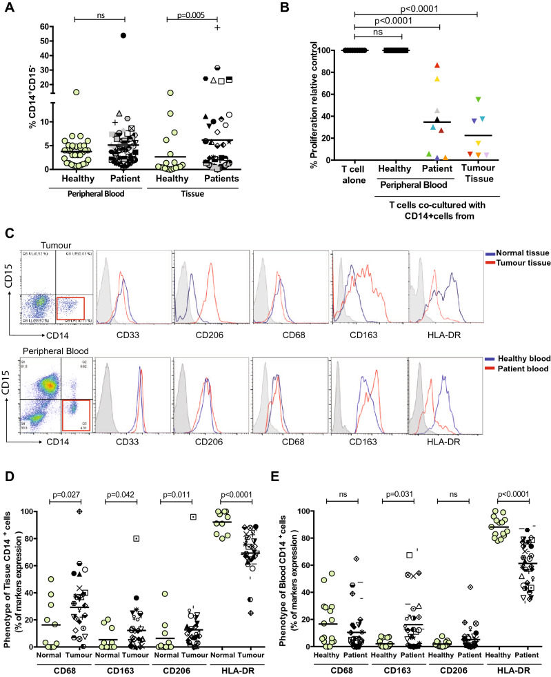
Background: Monocytic myeloid-derived suppressor cells (M-MDSCs) are significantly expanded in the blood of colorectal cancer (CRC) patients. However, their presence and underlying mechanisms in the tumour microenvironment of CRC have not been examined in detail.

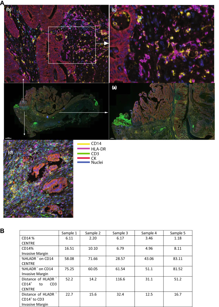
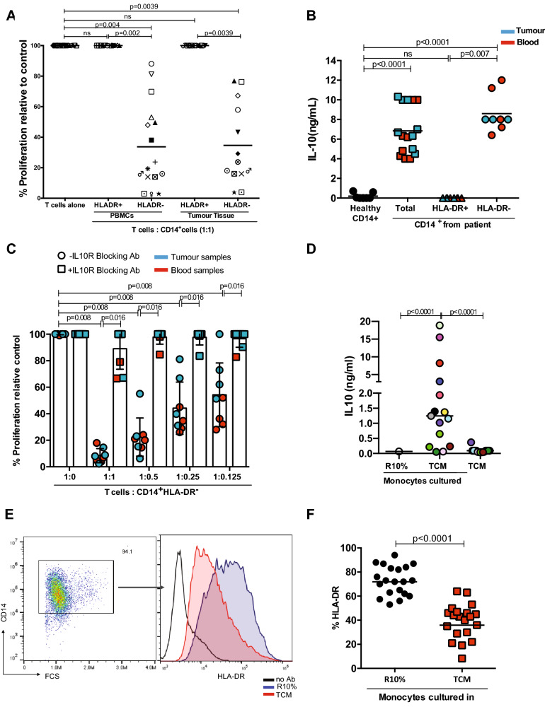
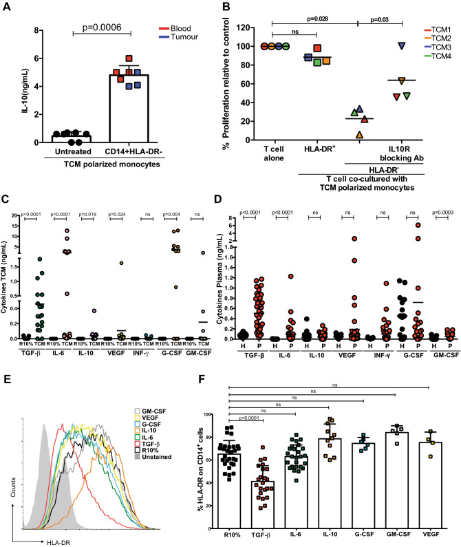
Methods: Tumour tissues and peripheral blood from CRC patients were analysed for the presence of M-MDSCs. The mechanisms of suppression were analysed by blocking pathways by which MDSCs abrogate T cell proliferation. Co-culture of CRC cells with monocytes were performed with and without cytokine blocking antibodies to determine the mechanism by which CRC cells polarise monocytes. Multi-spectral IHC was used to demonstrate the intra-tumoral location of M-MDSCs.

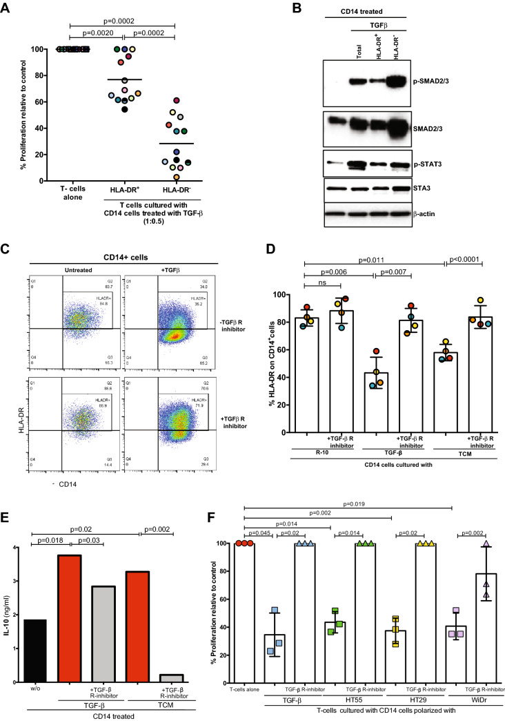
Results: Tumour tissues and blood of CRC patients contain M-MDSCs which inhibit T cell proliferation. Whilst inhibition of arginase and nitric oxide synthase 2 fail to rescue T cell proliferation, blockade of IL-10 released by these HLA-DR- cells abrogates the suppresivity of M-MDSCs. Tumour conditioned media (TCM) significantly reduces HLA-DR expression, increases IL-10 release from monocytes and causes them to become suppressive. TGF-β is highly expressed in the TCM and accumulates in the plasma. TGF-β reduces HLA-DR expression and drives monocyte immunosuppressivity. The invasive margin of CRC is enriched in CD14+ HLA-DR- cells in close proximity to T cells.

Conclusions: Our study demonstrates the cross-talk between CRC cells, M-MDSCs and T cells. Characterisation of CRC M-MDSCs point to therapeutic avenues to target these cells in addition to TGF-β blockade.

Keywords: Colorectal cancer; IL-10; M-MDSCs; TGFβ; pSTAT3.


Conflict of interest statement

The authors have no disclosures and no competing interests in relation to this manuscript or work.

Figures

Fig. 1
Fig. 1
CD14+ cells in the blood and tumour suppress peripheral blood leukocyte proliferation in CRC patients a Increased frequency of CD14+ monocytes staining in the peripheral blood (n=53) and tissue (n=18) of colorectal cancer samples and healthy control (p=0.005). b Allogeneic T cell proliferation under anti-CD3/CD28 antibody stimulation is suppressed with the addition of CD14+ cells from tissue (n=7) and the blood (n=9) of CRC patients, as measured by 3H-Tymidine uptake, compared to CD14+cells from healthy donors (p<0.0001). c Phenotypic analysis of CD14+ cells from blood and tissue of representative CRC patient. Flow analysis of CD14+ cells shows an increased expression of CD206,CD68, CD163 markers in the tumour tissue d and not in the blood e of CRC patients, instead and a significant down regulation of HLA-DR expression is measured in both the compartments compared healthy control
Fig. 2
Fig. 2
Tumour localization of CD14+ myeloid cells in colorectal cancer. A) Representative multispectral immunohistochemical staining of colorectal cancer section. The squares in the middle right panel and magnified in (a) indicates the area of colon analysed with higher magnification where arrows indicate an increase in magnification of the specified area - upper squares, centre of tumour and lower squares, invasive margin: the magnified invasive margin image is shown in (d) and those of the tumour centre are shown in (b and c). CD14: Yellow; HLA-DR: Magenta; CD3: Cyan/green; CK:Red; nuclei: Blue. B) Frequency of CD14+ cells as a percentage of total cells in colorectal cancer tissue at the tumour centre and invasive margins, the percentage of DR–CD14+ cells and the distance of DR–CD14+ cells to CD3+ cells as determined by VECTRA spectral analysis (n=5)
Fig. 3
Fig. 3
CD14+HLA-DR- cells are the major immunosuppressive cells in CRC. a Allogeneic T cell proliferation under anti-CD3/CD28 antibody stimulation is suppressed with the addition of CD14+HLA-DR- cells from the blood (n=16) and the tissue (n=13) of CRC patients, as measured by 3HTymidine uptake. The CD14+HLA-DR+ population is not suppressive. The 1:1 ratio of T cells:CD14+HLA-DR- cells is shown. Each colour represents a CRC patient. b Significantly higher level of IL-10 was detected by ELISA in the supernatant of CD14+ (blood samples n=8, tumour tissue n=8)and CD14+HLA-DR- cells (blood samples n=5, tumour tissue n=3) compared healthy control. c IL-10 receptor blocking antibody (10μg/ml) inhibits the suppressive activity of CD14+HLA-DR- cells of CRC patients restoring T cell proliferation (n=10). d Colorectal tumour conditioned media (TCMs, n=15)drive the release of IL10 from healthy monocytes after 48h incubation. e A representative histogram of HLA-DR expression on monocytes after 48h incubation with TCM. f Down regulation of HLA-DR on monocytes cultured with 20 TCMs
Fig. 4
Fig. 4
Colorectal tumour conditioned media drives IL10 secretion. a Significantly higher level of IL-10 was detected by ELISA in the supernatant of CD14+HLA-DR- cells polarized by TCM (blood samples n=4, tumour tissue n=3) compared healthy control. b IL-10 receptor blocking antibody (10μg/ml) inhibits the suppressive activity of TCMs polarized CD14+HLA-DR- cells restoring T cell proliferation (n=4). Higher concentration of TGF-β was measured by ELISA in TCM (n=19) c and in the plasma d of colorectal patients (n=39 and healthy plasma n=10). e A representative histogram of HLA-DR expression on monocytes cultured with cytokines for 48h. f Down regulation of HLA-DR on monocytes cultured with cytokines for 48h was observed by Flow analysis (n=23 TGFβ, n=26 IL-6, n=11 IL-10, n=6 G-CSF, n=5 GM-CSF and n=4 VEGF)
Fig. 5
Fig. 5
TGF-β derived M-MDSC suppress T cell proliferation. a TGF-β polarized CD14+HLA-DRcells suppress T-cell proliferation. Less suppressive activity has shown from TGF-β polarized CD14+HLA-DR+ cells. The 1:0.5 ratio of T cells: myeloid cells shown (n=13). b High expression of p-SMAD and p-STAT3 was detected by Western blot in the CD14+HLA-DR- cells polarized by TGF-β.Representative of 4 experiments. c and d TGF-β receptor inhibitor inhibits the ability of TGF-β and TCMs to down regulate HLA-DR on polarized monocytes (n=4). e TGF- β receptor inhibitor inhibits the release of IL-10 from TGF-β and TCM polarized monocytes. Representative of 2 experiments. fTGF- β receptor inhibitor inhibits the suppressive activity of CD14+HLA-DR- cells polarized with TGF-β and CRC cell TCM (n=3)

References

    1. Bronte V, Brandau S, Chen SH, Colombo MP, Frey AB, Greten TF, et al. Recommendations for myeloid-derived suppressor cell nomenclature and characterization standards. Nat Comms. 2016;7:12150. doi: 10.1038/ncomms12150. - DOI - PMC - PubMed
    1. http://www.cancerresearchuk.org/health-professional/cancer-statistics/st...
    1. Kim K, Skora AD, Li Z, Liu Q, Tam AJ, Blosser RL, et al. Eradication of metastatic mouse cancers resistant to immune checkpoint blockade by suppression of myeloid-derived cells. Proc Natl Acad Sci U S A. 2014;111(32):11774–11779. doi: 10.1073/pnas.1410626111. - DOI - PMC - PubMed
    1. Highfill SL, Cui Y, Giles AJ, Smith JP, Zhang H, Morse E, et al. Disruption of CXCR2-mediated MDSC tumor trafficking enhances anti-PD1 efficacy. Sci Transl Med. 2014;6(237):23ra7673. doi: 10.1126/scitranslmed.3007974. - DOI - PMC - PubMed
    1. Duffy A, Zhao F, Haile L, Gamrekelashvili J, Fioravanti S, Ma C, et al. Comparative analysis of monocytic and granulocytic myeloid-derived suppressor cell subsets in patients with gastrointestinal malignancies. Cancer Immunol Immunother. 2013;62(2):299–307. doi: 10.1007/s00262-012-1332-3. - DOI - PMC - PubMed