. 2022 Feb;52(2):297-311.
doi: 10.1002/eji.202149331. Epub 2021 Nov 23.

Profiling the specificity of clonally expanded plasma cells during chronic viral infection by single-cell analysis

Affiliations

Profiling the specificity of clonally expanded plasma cells during chronic viral infection by single-cell analysis

Daniel Neumeier et al. Eur J Immunol. 2022 Feb.

Abstract

Plasma cells and their secreted antibodies play a central role in the long-term protection against chronic viral infection. However, due to experimental limitations, a comprehensive description of linked genotypic, phenotypic, and antibody repertoire features of plasma cells (gene expression, clonal frequency, virus specificity, and affinity) has been challenging to obtain. To address this, we performed single-cell transcriptome and antibody repertoire sequencing of the murine BM plasma cell population following chronic lymphocytic choriomeningitis virus infection. Our single-cell sequencing approach recovered full-length and paired heavy- and light-chain sequence information for thousands of plasma cells and enabled us to perform recombinant antibody expression and specificity screening. Antibody repertoire analysis revealed that, relative to protein immunization, chronic infection led to increased levels of clonal expansion, class-switching, and somatic variants. Furthermore, antibodies from the highly expanded and class-switched (IgG) plasma cells were found to be specific for multiple viral antigens and a subset of clones exhibited cross-reactivity to nonviral and autoantigens. Integrating single-cell transcriptome data with antibody specificity suggested that plasma cell transcriptional phenotype was correlated to viral antigen specificity. Our findings demonstrate that chronic viral infection can induce and sustain plasma cell clonal expansion, combined with significant somatic hypermutation, and can generate cross-reactive antibodies.

Keywords: LCMV; immune receptor repertoire sequencing; plasma cells; single-cell; viral infection.


Conflict of interest statement

Authors declare that there are no conflict of interest.

Figures
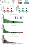
Figure 1
Figure 1
Single‐cell immune repertoire sequencing reveals clonal expansion and class‐switching in the BM plasma cell repertoire following chronic viral infection. (A) Experimental overview of chronic LCMV infection, BM plasma cell isolation, and single‐cell sequencing. (B) Number of cells per isotype for each infected or immunized mouse. Only cells containing exactly one variable heavy (VH) and variable light (VL) chain were considered. Colors correspond to isotype. (C) Number of clones per isotype for each infected or immunized mouse. Clones were determined by grouping those B cells containing identical CDRH3 + CDRL3 AA sequences. The isotype was determined as the isotype corresponding to the majority of cells within one clone. Color corresponds to isotype. (D) Clonal expansion for the top 50 most expanded clones of the BM plasma cells for each infected or immunized mouse. Clones were determined by grouping those B cells containing identical CDRH3 + CDRL3 AA sequences. Color corresponds to isotype.
Figure 2
Figure 2
Clonal expansion and number of antibody variants in the IgG BM plasma cell repertoire following chronic viral infection as compared to protein immunizations. (A) Clonal expansion of the BM plasma cells (PC) separated by isotype for a single mouse following chronic LCMV infection. The number of distinct cell barcodes belonging to the top 30 clones is shown. Clone was determined by grouping those B cells containing identical CDRH3 + CDRL3 AA sequences. Only cells containing exactly one variable heavy (VH) and variable light (VL) chain AA sequence were considered. The isotype was determined as the isotype corresponding to the majority of cells within one clone. Color corresponds to isotype. (B) Normalized clonal expansion separated by isotype for each of the infected or immunized mice. The number of cell barcodes per clone was divided by the total number of cells in each repertoire. (C) Number of cells per clone for the top 30 clones separated by isotype for each of the infected or immunized mice. (D) Quantity of AA variants within each clone separated by isotype for each of the infected or immunized mice. AA variants were determined by quantifying the number of unique full‐length VH + VL AA sequence variants within each clone. Clone was determined by grouping those B cells containing identical CDRH3 + CDRL3 AA sequences. (E) Relationship between the number of unique AA variants and the number of cell barcodes for the top 30 clones separated by isotype majority following chronic LCMV infection.
Figure 3
Figure 3
Transcriptional heterogeneity of BM PCs from LCMV‐infected or TNFR2‐immunized mice. (A) Uniform manifold approximation projection (UMAP)‐based total gene expression for BM plasma cell (BM PC) repertoire following either LCMV infection or TNFR2 immunization. Each point corresponds to a cell and color corresponds to the transcriptional cluster. (B) The fraction of cells belonging to each transcriptional cluster from the BM PC repertoire following either LCMV infection or TNFR2 immunization. (C) Differentially expressed genes between BM PCs following LCMV infection and TNFR2 immunization. The order of genes (from top to bottom) corresponds to the highest average log‐fold change. All genes displayed have adjusted p values < 0.01. (D) UMAP displaying isotype distribution of BM PCs following either LCMV infection or TNFR2 immunization. Each point corresponds to a cell and color indicates isotype based on VDJ sequencing data. € Cluster membership separated by isotype for the BM PCs following LCMV infection. Color indicates the transcriptional cluster from panel A. (F) Cluster membership separated by isotype for the BM PCs following TNFR2 immunization. Color indicates the transcriptional cluster from panel A. (G) UMAP displaying clonal expansion for the BM PCs following either LCMV infection or TNFR2 immunization. “Expanded” corresponds to those clones supported by two or more unique cell barcodes. Clone was determined by grouping those B cells containing identical CDRH3 + CDRL3 AA sequences. (H) Cluster membership separated by clonal expansion for the BM PCs following either LCMV infection or TNFR2 immunization. Color indicates the transcriptional cluster from panel A.
Figure 4
Figure 4
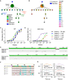
Clonally expanded plasma cells are virus‐specific and potentially autoreactive. (A) The ELISA signal of duplicate measurements at 450 nm is shown relative to a negative background control (red dotted line indicates background level). Clone was determined by grouping those B cells containing identical CDRH3 + CDRL3 AA sequences. Only cells containing exactly one variable heavy (VH) and variable light (VL) chain were considered. The isotype was determined as the isotype corresponding to the majority of cells within one clone. For each clone, the antibody variant (combined VH+VL nucleotide sequence) supported by the most unique cell barcodes was selected to be expressed. Clones 13, 24, 26, and 29 did not express in our hybridoma system and were, therefore, excluded from the ELISA. (B) Neutralization potential of the GPC‐ and lysate‐binding clones normalized by wells without antibody. (C) Flow cytometry confirmed GPC‐binding inferred by ELISA of antibody expressing hybridomas. (D) Mutational network of the IgG clone binding MC57G lysate. Nodes represent unique antibody variants (combined VH + VL nucleotide sequence) and edges demonstrate sequences with the smallest separation calculated by edit distance. Node color corresponds to transcriptional cluster from 3A. The size and label of the nodes indicate how many cells express each full‐length antibody variant. Clone was determined by grouping those B cells containing identical CDRH3 + CDRL3 AA sequences. Only cells containing exactly one variable heavy (VH) and variable light (VL) chain were considered. The isotype was determined as the isotype corresponding to the majority of cells within one clone. The germline node represents the unmutated reference sequence determined by 10× Genomics cell ranger. (E) Differentially expressed genes between LCMV NP, GPC binders, and MC57G lysate binders. Those labeled and colored points correspond to those genes with an adjusted p value of < 0.01. G. ELISA on lysate from various tissues of naive C57BL/6 mice. Error bars indicate the SE of mean.
Figure 5
Figure 5
Repertoire and transcriptome profile of LCMV‐specific PCs. (A) Sequence logo plots of the confirmed GPC, NP, and MC57G lysate binders. (B) Isotype distribution for the confirmed GPC, NP, and MC57G lysate binders. (C) Location of the confirmed GPC, NP, and MC57G lysate binders on the UMAP. (D) Transcriptome distribution for the confirmed GPC, NP, and MC57G lysate binders. Color indicates the transcriptional cluster from 3A.
Figure 6
Figure 6
Virus‐specific somatic variants present in the BM PC repertoire are cross‐reactive. (A,B) Mutational network of the NP‐specific, second most expanded IgG clone. Nodes represent unique antibody variants (combined VH+VL nucleotide sequence) and edges demonstrate sequences with the smallest separation calculated by edit distance. Node color corresponds to either isotype (A) or transcriptional cluster (B). The size and label of the nodes indicate how many cells express each full‐length antibody variant. Clone was determined by grouping those B cells containing identical CDRH3 + CDRL3 AA sequences. Only cells containing exactly one variable heavy (VH) and variable light (VL) chain were considered. The isotype was determined as the isotype corresponding to the majority of cells within one clone. Variant labels indicate those full‐length antibodies which were recombinantly expressed. The germline node represents the unmutated reference sequence determined by 10× Genomics cellranger. (C) NP ELISA for the four variants from the second most expanded IgG clone. (D) DNP‐OVA ELISA for the four variants from the second most expanded IgG clone. (E) VH and VL nucleotide alignments of the four variants from the second most expanded IgG clone. (F) VH amino acid alignment highlighting mutations in the CDRH2. Colored graphs correspond to isoelectric point (PI). (G) Affinity measurements (kd ) against LCMV NP for the four variants from the second most expanded IgG clone in molar (M) concentrations.

References

    1. Bootz, A. , Karbach, A. , Spindler, J. , Kropff, B. , Reuter, N. , Sticht, H. , Winkler, T. H. et al., Protective capacity of neutralizing and non‐neutralizing antibodies against glycoprotein B of cytomegalovirus. PLoS Pathog. 2017. 13: e1006601. - PMC - PubMed
    1. Burton, D. R. and Hangartner, L. , Broadly neutralizing antibodies to HIV and their role in vaccine design. Annu. Rev. Immunol. 2016. 34: 635–659. - PMC - PubMed
    1. Greczmiel, U. , Kräutler, N. J. , Pedrioli, A. , Bartsch, I. , Agnellini, P. , Bedenikovic, G. , Harker, J. et al., Sustained T follicular helper cell response is essential for control of chronic viral infection. Sci. Immunol. 2017. 2: eaam8686. - PubMed
    1. Nguyen, D. C. , Joyner, C. J. , Sanz, I. and Lee, F. E. ‐H. , Factors affecting early antibody secreting cell maturation into long‐lived plasma cells. Front. Immunol. 2019. 10: 2138. - PMC - PubMed
    1. Battegay, M. , Moskophidis, D. , Waldner, H. , Bründler, M. A. , Fung‐Leung, W. P. , Mak, T. W. , Hengartner, H. et al., Impairment and delay of neutralizing antiviral antibody responses by virus‐specific cytotoxic T cells. J. Immunol. 1993. 151: 5408–5415. - PubMed

Publication types

MeSH terms

Substances