

Pleural effusion volume in patients with acute pancreatitis: a retrospective study from three acute pancreatitis centers
- PMID: 34727802
- PMCID: PMC8567956
- DOI: 10.1080/07853890.2021.1998594
Pleural effusion volume in patients with acute pancreatitis: a retrospective study from three acute pancreatitis centers
Abstract
Objective: To assess the value of pleural effusion volume (PEV) quantified on chest computed tomography (CT) in patients with early stage acute pancreatitis (AP).
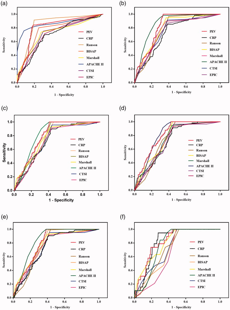
Methods: Data of PEV, and C-reactive protein (CRP) levels as well as Ranson, bedside index of severity in acute pancreatitis (BISAP), Marshall, acute physiology and chronic health evaluation II (APACHE II), CT severity index (CTSI), and extra-pancreatic inflammation on computed tomography (EPIC) scores in patients with AP were collected. Duration of hospitalization, severity of AP, infection, procedure, intensive care unit (ICU) admission, organ failure, or death were included as the outcome parameters.
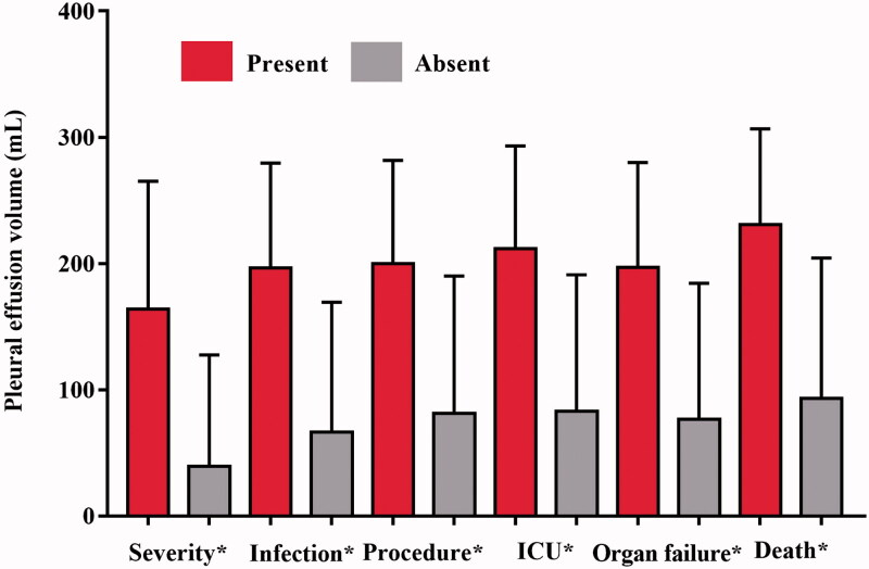
Results: In 465 patients, the mean PEV was 98.8 ± 113.2 mL. PEV showed strong and significant correlations with the CRP levels, duration of hospitalization as well as the Ranson, BISAP, Marshall, APACHE II, CTSI, and EPIC scores (p < .05). PEV demonstrated significant accuracy in predicting severity, infection, procedure, ICU admission, organ failure, and death (p < .05).
Conclusion: PEV quantified on chest CT positively associated with the duration of hospitalization, CRP levels, Ranson, BISAP, Marshall, APACHE II, CTSI, and EPIC scores. It can be a reliable radiologic biomarker in predicting severity and clinical outcomes of AP.KEY MESSAGESPleural effusion is a common chest finding in patients with acute pancreatitis.Pleural effusion volume quantified on chest CT examination positively associated with the duration of hospitalization, CRP level, as well as Ranson, BISAP, Marshall, APACHE II, CTSI, and EPIC scoring systems.Pleural effusion volume can be a reliable radiologic biomarker in the prediction of severity and clinical outcomes of acute pancreatitis.
Keywords: Pleural effusion; acute pancreatitis; computed tomography.
Conflict of interest statement
The authors have no conflicts of interest to declare.
Figures
References
-
- Lankisch PG, Apte M, Banks PA.. Acute pancreatitis. Lancet. 2015;386(9988):85–96. - PubMed
-
- Forsmark CE, Vege SS, Wilcox CM.. Acute pancreatitis. N Engl J Med. 2016;375(20):1972–1981. - PubMed
-
- Majidi S, Golembioski A, Wilson SL, et al. Acute pancreatitis: etiology, pathology, diagnosis, and treatment. South Med J. 2017;110(11):727–732. - PubMed
-
- Zhu Y, Pan X, Zeng H, et al. A study on the etiology, severity, and mortality of 3260 patients with acute pancreatitis according to the revised Atlanta classification in Jiangxi, China over an 8-year period. Pancreas. 2017;46(4):504–509. - PubMed
Publication types
MeSH terms
Substances
LinkOut - more resources
Full Text Sources
Medical
Research Materials
Miscellaneous