Review
. 2022 Feb;162(2):415-430.
doi: 10.1053/j.gastro.2021.10.036. Epub 2021 Oct 30.

Cellular Plasticity, Reprogramming, and Regeneration: Metaplasia in the Stomach and Beyond

Affiliations
Review

Cellular Plasticity, Reprogramming, and Regeneration: Metaplasia in the Stomach and Beyond

James R Goldenring et al. Gastroenterology. 2022 Feb.

Abstract

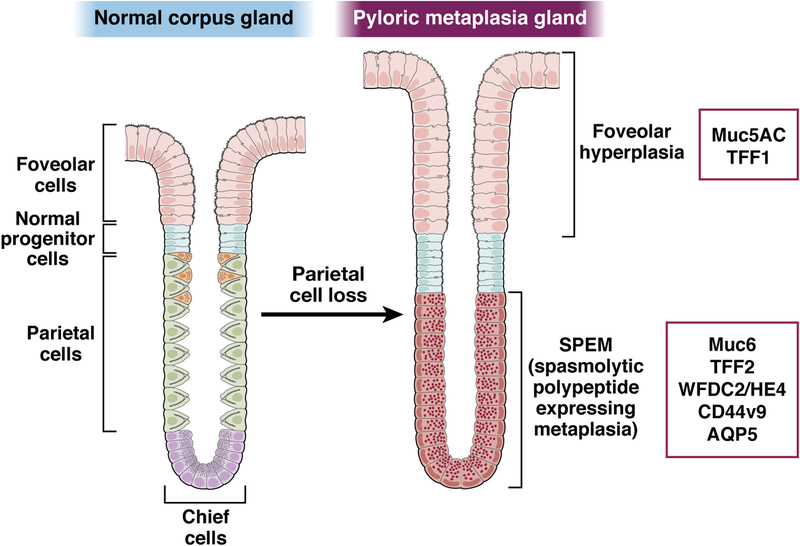
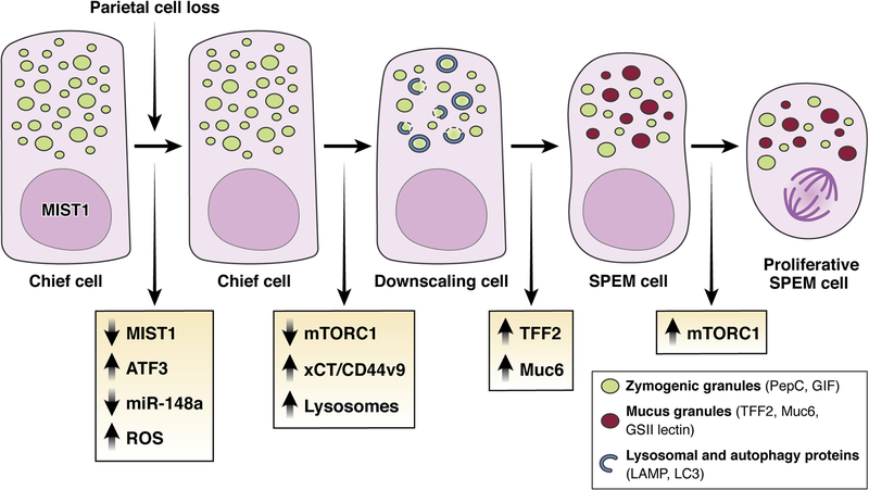
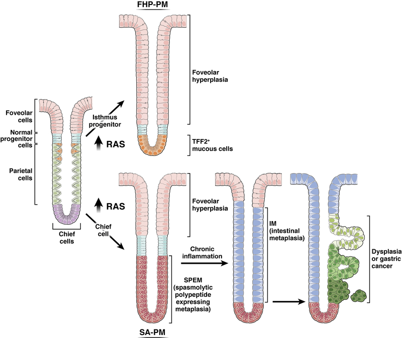
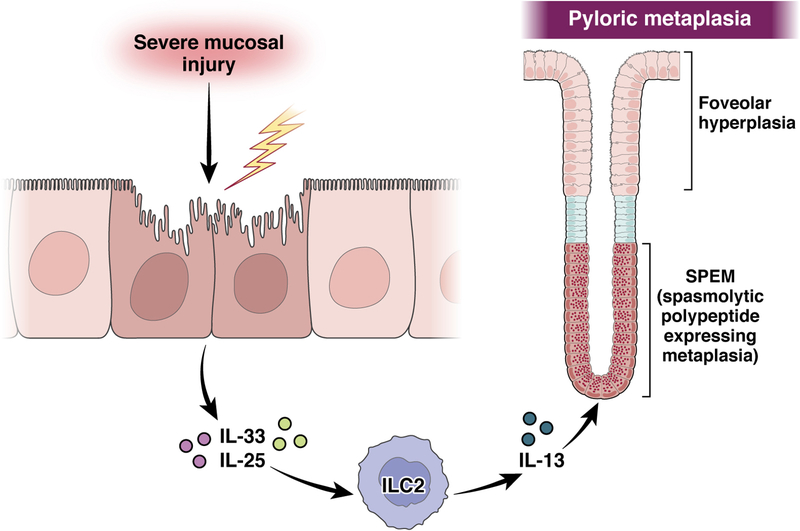
The mucosa of the body of the stomach (ie, the gastric corpus) uses 2 overlapping, depth-dependent mechanisms to respond to injury. Superficial injury heals via surface cells with histopathologic changes like foveolar hyperplasia. Deeper, usually chronic, injury/inflammation, most frequently induced by the carcinogenic bacteria Helicobacter pylori, elicits glandular histopathologic alterations, initially manifesting as pyloric (also known as pseudopyloric) metaplasia. In this pyloric metaplasia, corpus glands become antrum (pylorus)-like with loss of acid-secreting parietal cells (atrophic gastritis), expansion of foveolar cells, and reprogramming of digestive enzyme-secreting chief cells into deep antral gland-like mucous cells. After acute parietal cell loss, chief cells can reprogram through an orderly stepwise progression (paligenosis) initiated by interleukin-13-secreting innate lymphoid cells (ILC2s). First, massive lysosomal activation helps mitigate reactive oxygen species and remove damaged organelles. Second, mucus and wound-healing proteins (eg, TFF2) and other transcriptional alterations are induced, at which point the reprogrammed chief cells are recognized as mucus-secreting spasmolytic polypeptide-expressing metaplasia cells. In chronic severe injury, glands with pyloric metaplasia can harbor both actively proliferating spasmolytic polypeptide-expressing metaplasia cells and eventually intestine-like cells. Gastric glands with such lineage confusion (mixed incomplete intestinal metaplasia and proliferative spasmolytic polypeptide-expressing metaplasia) may be at particular risk for progression to dysplasia and cancer. A pyloric-like pattern of metaplasia after injury also occurs in other gastrointestinal organs including esophagus, pancreas, and intestines, and the paligenosis program itself seems broadly conserved across tissues and species. Here we discuss aspects of metaplasia in stomach, incorporating data derived from animal models and work on human cells and tissues in correlation with diagnostic and clinical implications.


Conflict of interest statement

The authors have no conflicts of interest.

Figures

Figure 1:
Figure 1:. Markers of pyloric metaplasia.
Pyloric metaplasia evolves in the corpus of the stomach following parietal cell loss with the establishment of SPEM cells at the base of glands and often foveolar hyperplasia luminal to the SPEM lineages. Foveolar hyperplasia is marked by expansion of cells expressing Muc5AC and TFF1. SPEM is identified by cells expressing chief cell markers in low abundance plus induced AQP5, MUC6, TFF2, WFDC2/HE4 and CD44v9.
Figure 2:
Figure 2:. Evolution of SPEM through transdifferentiation/paligenosis.
Following parietal cell loss, chief cells rapidly down-regulate expression of the chief-cell-architecture master regulating transcription factor Mist1 and the micro-RNA miR-148a. mTORC1 is downregulated, and reactive oxygen species (ROS) increase. Loss of MIST1, decrease of mTORC1, and ROS facilitate expansion of lysosomal elements to remove damaged ROS-producing organelles and consume excess zymogen granules. Next, cellular ROS-adaptive mechanisms such as xCT/CD44v9 are increased. Management of ROS and downscaling of zymogen granules are required before the next stage: up-regulation of TFF2 and MUC6 expression and the assembly of mucus-secreting granules that define the reprogramming into a SPEM cell phenotype. Continued damage and inflammation can promote the progression of SPEM into a more proliferative lineage and transition to this stage requires re-induction of mTORC1.
Figure 3:
Figure 3:. The divergent effects of Ras activation in isthmal progenitor cells and chief cells establish two types of pyloric metaplasia.
Increases in Ras activation in isthmal progenitor cells in transgenic mice and in the setting of Ménétrier’s disease leads to massive foveolar hyperplasia (foveolar hyperplasia predominant pyloric metaplasia, FHP-PM) with preferential differentiation of foveolar cells, and to a lesser extent mucous neck cells, over parietal cells. In contrast, activation of Ras in chief cells leads initially to the development of SPEM from reprogramming of chief cells (SPEM-associated pyloric metaplasia, SA-PM). Continuously active Ras expression can lead to intestinal metaplasia and dysplasia, establishing the full range of pre-neoplastic lineages associated with the development of intestinal type gastric cancer. Note: the intestinal metaplasia depicted here would be of the “incomplete type.” The figure is not meant to show definitively which exact cells are the ones fueling the expansion of dysplastic/neoplastic cells, but evidence does point to lineages at the interface of intestinal metaplasia arising from pyloric metaplasia/SPEM as the figure depicts.
Figure 4:
Figure 4:. Intrinsic immune cells coordinate the initiation of SPEM.
Severe damage in the corpus mucosa leads to the release of IL-33 from surface mucous cells and IL-25 from tuft cells, both of which stimulate the release of IL-13 from ILC2s. IL-13 in turn is required for initiation of the reprogramming of chief cells into SPEM cells and the development of pyloric metaplasia.

References

    1. Choi E, Roland JT, Barlow BJ, et al. Cell lineage distribution atlas of the human stomach reveals heterogeneous gland populations in the gastric antrum. Gut 2014;63:1711–20. - PMC - PubMed
    1. Engevik AC, Kaji I, Goldenring JR. The Physiology of the Gastric Parietal Cell. Physiol Rev 2020;100:573–602. - PMC - PubMed
    1. Saenz JB, Mills JC. Acid and the basis for cellular plasticity and reprogramming in gastric repair and cancer. Nat Rev Gastroenterol Hepatol 2018;15:257–273. - PMC - PubMed
    1. Aihara E, Medina-Candelaria NM, Hanyu H, et al. Cell injury triggers actin polymerization to initiate epithelial restitution. J Cell Sci 2018;131. - PMC - PubMed
    1. Aihara E, Hentz CL, Korman AM, et al. In vivo epithelial wound repair requires mobilization of endogenous intracellular and extracellular calcium. J Biol Chem 2013;288:33585–97. - PMC - PubMed

Publication types