. 2021 Dec 15:245:118681.
doi: 10.1016/j.neuroimage.2021.118681. Epub 2021 Oct 30.

Age-related decline in cortical inhibitory tone strengthens motor memory

Affiliations

Age-related decline in cortical inhibitory tone strengthens motor memory

Pierre Petitet et al. Neuroimage. .

Abstract

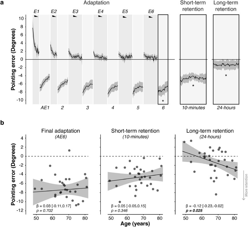
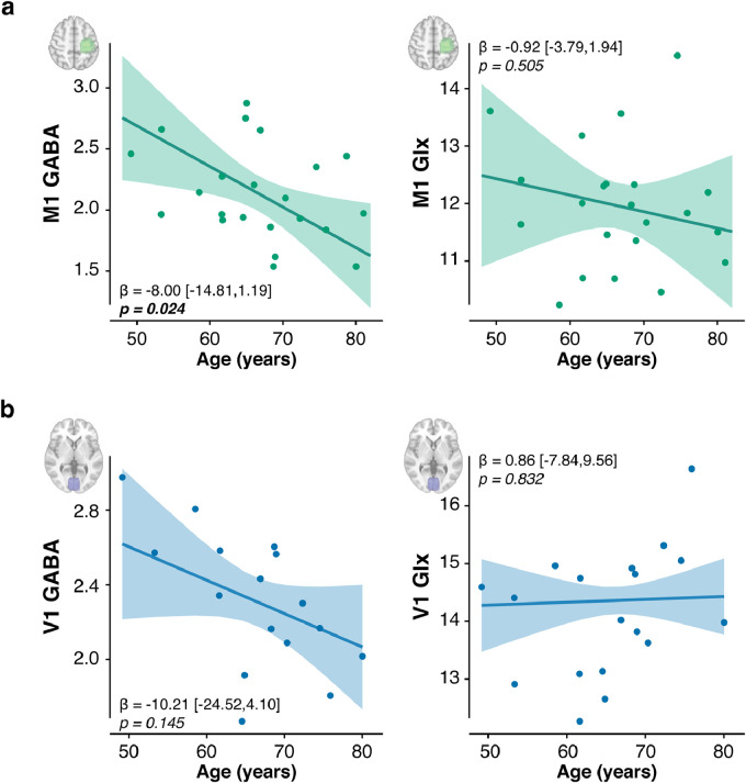
Ageing disrupts the finely tuned excitation/inhibition balance (E:I) across cortex via a natural decline in inhibitory tone (γ-amino butyric acid, GABA), causing functional decrements. However, in young adults, experimentally lowering GABA in sensorimotor cortex enhances a specific domain of sensorimotor function: adaptation memory. Here, we tested the hypothesis that as sensorimotor cortical GABA declines naturally with age, adaptation memory would increase, and the former would explain the latter. Results confirmed this prediction. To probe causality, we used brain stimulation to further lower sensorimotor cortical GABA during adaptation. Across individuals, how stimulation changed memory depended on sensorimotor cortical E:I. In those with low E:I, stimulation increased memory; in those with high E:I stimulation reduced memory. Thus, we identified a form of motor memory that is naturally strengthened by age, depends causally on sensorimotor cortex neurochemistry, and may be a potent target for motor skill preservation strategies in healthy ageing and neurorehabilitation.

Keywords: Ageing; Excitation:inhibition ratio; Sensorimotor adaptation.


Conflict of interest statement

Declaration of Competing Interest The authors declare no competing interests.

Figures

None
Graphical abstract
Fig. 1
Fig. 1
Long-term retention of prism adaptation is higher in older adults. a. Group mean pointing errors expressed as change from baseline accuracy (y=0). Positive y-axis values are rightward errors (i.e. in the direction of the prismatic shift), negative leftward. Error bands indicate s.e.m. Black wedges indicate blocks in which prisms were worn. During right-shifting prism exposure (E1-6), visual feedback enabled participants to correct their rightward pointing errors across trials. Consequent leftward AE was measured in intervening blocks without visual feedback throughout adaptation (AE1-6). After-effect retention was measured post-adaptation after a short (10 min) and long (24 h) interval. There was significant retention at both time points. Asterisks indicate significant one-sample t-tests for the block-averaged AE against zero (p<0.05). b. Age had no effect on the AE magnitude acquired by the end of adaptation (block AE6), nor on short-term retention (10 min). The key finding was that older adults showed significantly greater long-term retention (24-hours). Error bands represent the 95% Confidence Intervals. Full statistics are in Tables S3 & S4.
Fig. 2
Fig. 2
Motor cortical inhibitory tone is lower in older adults. a. The concentration of GABA but not Glutamix (Glutamate + Glutamine, Glx) was associated negatively with age in the left sensorimotor cortex (labelled "M1"). b. There was no significant association between age and neurochemical concentration in occipital cortex (labelled "V1"). For each voxel and neurotransmitter, plotted relationships control for the fraction of grey matter and white matter, and the other neurotransmitter. Absolute concentrations are expressed in arbitrary units. Error bands represent the 95% Confidence Intervals. Full statistical details are in Table S5.
Fig. 3
Fig. 3
Lower motor cortical inhibitory tone is associated with greater long-term retention. Plot shows relationships between brain chemistry and the magnitude of prism after-effect retained 24 h after adaptation. Negative values on the y-axis indicate retention. a. Sensorimotor cortex ("M1") Across individuals, lower GABA was associated with greater retention. There was no relationship with Glx (Glutamate + Glutamine). b. Occipital cortex ("V1") There was no relationship between GABA or Glx and 24-hour retention. For each voxel and neurotransmitter, relationships control for the fraction of grey matter and white matter, and the other neurotransmitter. Absolute concentrations are expressed in arbitrary units. Error bands represent the 95% Confidence Intervals. Full statistics details are in Table S6.
Fig. 4
Fig. 4
Adaptation memory is stronger in older age owing to the decline in motor cortical inhibitory tone. A mediation model tested whether M1 neurochemistry explained the relationship between age and retention. Consistent with our mechanistic hypothesis, GABA, but not Glx, mediated the positive relationship between age and 24-hour retention, explaining 64% of the variance. Standardised regression coefficients are reported next to the corresponding paths. Asterisks indicate significance (p<0.05). Full statistics: Table 6. Independent variable: Age. Dependent variable: AE 24-hours post-adaptation. Mediators: M1 GABA and Glx (controlling for grey and white matter tissue fractions).
Fig. 5
Fig. 5
On average across older adults excitatory stimulation of M1 during adaptation did not increase retention. Timecourse of pointing errors for the same behavioural paradigm and graph conventions as in Fig. 1, except that stimulation (anodal or sham tDCS) was applied to left M1 throughout the adaptation phase. Errors are normalised to baseline (pre-adaptation) accuracy. Negative values on the y-axis indicate prism after-effects. Error bands indicate s.e.m. After excitatory stimulation of M1 during adaptation, retention increased numerically but not significantly, contrary to our previous findings in young adults, but consistent with our expectations in this cohort of older adults.
Fig. 6
Fig. 6
How stimulation changes memory depends on motor cortical E:I. a. Individuals’ M1 E:I (Glx:GABA) is plotted against the stimulation effect (anodal - sham difference in normalised pointing error at 24-hour retention). On the y-axis, negative values indicate greater retention with anodal tDCS compared to sham. Positive values indicate the opposite. Across individuals, stimulation enhanced retention in those with low E:I and impaired retention in those with high E:I. These data confirm the hypothesis that retention depends causally on M1 E:I. b. The schematic offers an explanatory account of the data in panel a. Under the assumption that stimulation increases E:I across the group, the distribution of induced memory change has an inverted U-shape. This suggests there is an optimal range of E:I within which retention is maximal. The optimum differs across individuals. By increasing E:I, stimulation moves individuals with low E:I towards maximum, increasing retention. But for individuals with high E:I, who are close to maximum, stimulation exceeds the optimum and so retention becomes impaired. c. A moderation analysis confirmed that how stimulation changed memory varied as a function of M1 E:I (Glx:GABA × tDCS : t1419=2.40, p=0.017). For visualisation purposes, this interaction is illustrated using a median split on the M1 E:I data. The data (top) and model fit (bottom) are plotted separately for individuals with low versus high M1 E:I, and show opposing effects of excitatory stimulation on adaptation memory that depend on individuals’ M1 E:I. Error bands in panel a represent the 95% Confidence Intervals; error bands in panels c and d represent standard error of the mean.

References

    1. Anguera J.A., Reuter-Lorenz P.A., Willingham D.T., Seidler R.D. Failure to engage spatial working memory contributes to age-related declines in visuomotor learning. J. Cogn. Neurosci. 2011;23(1):11–25. doi: 10.1162/jocn.2010.21451. - DOI - PubMed
    1. Antonenko D., Schubert F., Bohm F., Ittermann B., Aydin S., Hayek D., Grittner U., Flöel A. tDCS-induced modulation of GABA levels and resting-state functional connectivity in older adults. J. Neurosci. 2017;37(15):4065–4073. doi: 10.1523/JNEUROSCI.0079-17.2017. - DOI - PMC - PubMed
    2. https://www.jneurosci.org/content/37/15/4065

    1. Bachtiar V., Near J., Johansen-Berg H., Stagg C.J. Modulation of GABA and resting state functional connectivity by transcranial direct current stimulation. eLife. 2015;4:e08789. doi: 10.7554/eLife.08789. - DOI - PMC - PubMed
    1. Barron H., Vogels T., Emir U., Makin T., O’Shea J., Clare S., Jbabdi S., Dolan R., Behrens T. Unmasking latent inhibitory connections in human cortex to reveal dormant cortical memories. Neuron. 2016;90(1):191–203. doi: 10.1016/j.neuron.2016.02.031. - DOI - PMC - PubMed
    2. https://www.sciencedirect.com/science/article/pii/S0896627316001689

    1. Bedard A.-C., Nichols S., Barbosa J.A., Schachar R., Logan G.D., Tannock R. The development of selective inhibitory control across the life span. Dev. Neuropsychol. 2002;21(1):93–111. doi: 10.1207/S15326942DN2101_5. - DOI - PubMed

Publication types

Substances