

Gray Lesions of the Breast and its Diagnostic Significance: A Retrospective Study from Rural India
- PMID: 34729352
- PMCID: PMC8507519
- DOI: 10.4103/JMAU.JMAU_19_20
Gray Lesions of the Breast and its Diagnostic Significance: A Retrospective Study from Rural India
Abstract
Background: Breast lesions extend from benign to malignant ones. The National Cancer Institute recommended categories for the diagnosis of breast cytology. There are some lesions in the breast which are called intermediate or gray lesions. It includes C3 (atypical, probably benign) and C4 (suspicious, favor malignant) which needs to be evaluated.
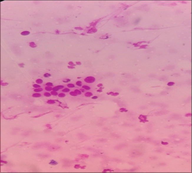
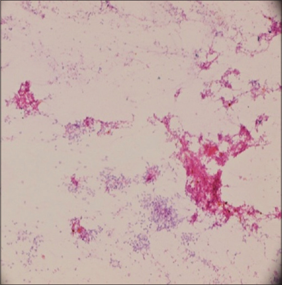
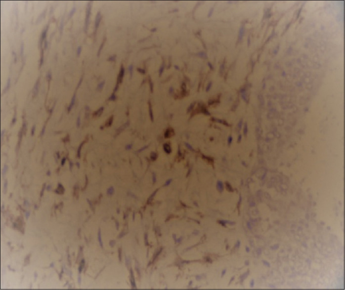
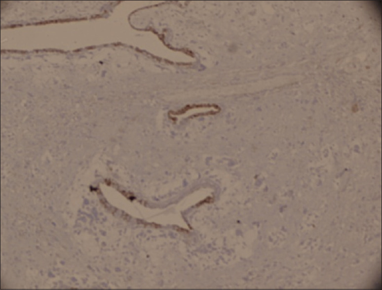
Materials and methods: This study was conducted in the Department of Pathology, Uttar Pradesh University of Medical Sciences, Saifai, Etawah (Uttar Pradesh). Fine-needle aspiration cytopathology (FNAC) was the diagnostic tool. The present study was undertaken to determine the gray lesions of the breast and its correlation with histopathology and other associated parameters. Immunohistochemistry was applied where ever necessary. One hundred and fifty one cases of gray lesions of the breast were included.
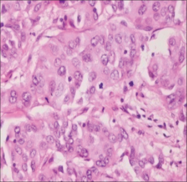
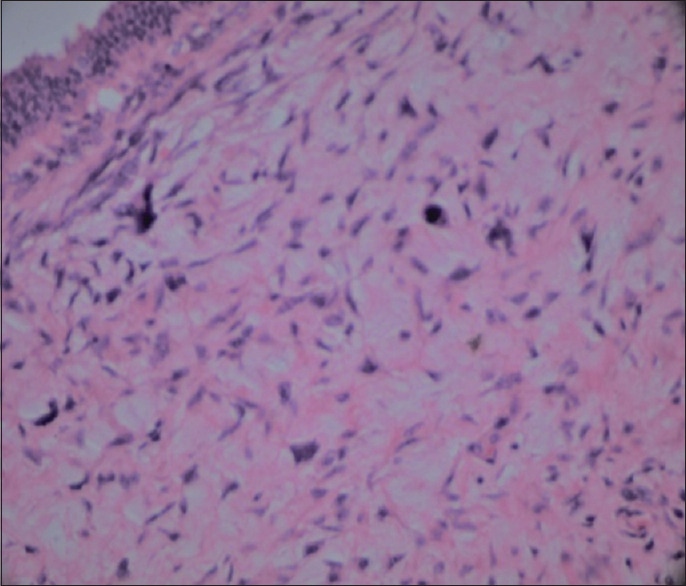
Results: C3 was seen in 85 (56.29%) and C4 in 66 (43.70%) patients. The maximum number of patients was of 31-40, (33.77%) years age group, the youngest patient was 12-year-old female, whereas the oldest was 86 years male. Histopathology evaluation confirmed malignancy in 35 (23.17%) cases, and infiltrating ductal carcinoma was the frequent malignancy (24 [68.5%]). Sensitivity, specificity, positive predictive value, and negative predictive value of C4 category for the diagnosis of malignancy were, respectively, 81.48%, 50%, 68.7%, and 64.2%.
Conclusion: FNAC is an excellent diagnostic tool. It has some limitations, especially with the gray lesions, which may lead to miss interpretation in diagnosis, so a scope of mistake to the cytopathologist is always there. These lesions need to be evaluated because of the risk of malignancy. However, gray lesions can be reduced by cytology followed by histopathology examination along with ancillary radiological investigations such as mammography and ultrasonography.
Keywords: Breast; fine needle aspiration cytopathology (FNAC); gray lesions; histopathology.
Copyright: © 2021 Journal of Microscopy and Ultrastructure.
Conflict of interest statement
There are no conflicts of interest.
Figures
References
-
- Moore KL, Persaud TV. Philadelphia, PA: W.B. Saunders Co; 1998. The integumentary system. In: The Developing Human: Clinically Oriented Embryology. Textbook of Embryology; pp. 513–30.
-
- Mandal AK, Choudhury S. Textbook of Pathology. 1st ed. New Delhi, India: Avichal Publishers; 2010. The breast; p. 663.
-
- Madan M, Sharma M, Piplani S, Manjari M, Sharma N, Goyal S. Evaluation of fine needle aspiration cytology in the diagnosis of suspiciou/gray zone lesions in breast lesions and its histopathological correlation. Ann Pathol Lab Med. 2016;3:573–6.
-
- Ferlay J, Soerjomataram I, Dikshit R. Cancer incidence and mortility worldwide: sources, methods and major patterns in GLOBO CAN 2012. Int J Cancer. 2015;136:E359–86. - PubMed
LinkOut - more resources
Full Text Sources
Miscellaneous