. 2021 Oct 10;3(1):vdab149.
doi: 10.1093/noajnl/vdab149. eCollection 2021 Jan-Dec.

Intraoperative DNA methylation classification of brain tumors impacts neurosurgical strategy

Affiliations

Intraoperative DNA methylation classification of brain tumors impacts neurosurgical strategy

Luna Djirackor et al. Neurooncol Adv. .

Abstract

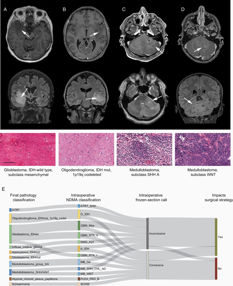
Background: Brain tumor surgery must balance the benefit of maximal resection against the risk of inflicting severe damage. The impact of increased resection is diagnosis-specific. However, the precise diagnosis is typically uncertain at surgery due to limitations of imaging and intraoperative histomorphological methods. Novel and accurate strategies for brain tumor classification are necessary to support personalized intraoperative neurosurgical treatment decisions. Here, we describe a fast and cost-efficient workflow for intraoperative classification of brain tumors based on DNA methylation profiles generated by low coverage nanopore sequencing and machine learning algorithms.

Methods: We evaluated 6 independent cohorts containing 105 patients, including 50 pediatric and 55 adult patients. Ultra-low coverage whole-genome sequencing was performed on nanopore flow cells. Data were analyzed using copy number variation and ad hoc random forest classifier for the genome-wide methylation-based classification of the tumor.

Results: Concordant classification was obtained between nanopore DNA methylation analysis and a full neuropathological evaluation in 93 of 105 (89%) cases. The analysis demonstrated correct diagnosis in 6/6 cases where frozen section evaluation was inconclusive. Results could be returned to the operating room at a median of 97 min (range 91-161 min). Precise classification of the tumor entity and subtype would have supported modification of the surgical strategy in 12 out of 20 patients evaluated intraoperatively.

Conclusion: Intraoperative nanopore sequencing combined with machine learning diagnostics was robust, sensitive, and rapid. This strategy allowed DNA methylation-based classification of the tumor to be returned to the surgeon within a timeframe that supports intraoperative decision making.

Keywords: DNA methylation; brain tumor; extent of resection; intraoperative diagnostics; nanopore.


Figures

Figure 1.
Figure 1.
Nanopore DNA methylation analysis (NDMA) gives a robust classification of brain tumors. Final neuropathological diagnosis (left) compared to NDMA diagnosis (center) and concordance of methods (right). “A” represents results from the 55 CNS tumors from adult cases reported in the study while “B” represents results from the 50 pediatric cases reported. In summary, NDMA was concordant with final neuropathological diagnosis in 93 of the 105 cases. Abbreviations: A, astrocytoma; ATRT, atypical teratoid rhabdoid tumor; CNS, central nervous system; CPH, craniopharyngioma; DMG, diffuse midline glioma; EPN, ependymoma; GBM, glioblastoma; MB, medulloblastoma; MNG, meningioma; O, oligodendroglioma; PLEX, plexus papilloma; PXA, pleomorphic xanthoastrocytoma; SCHW, schwannoma; SHH, sonic hedgehog.
Figure 2.
Figure 2.
Nanopore DNA methylation analysis (NDMA) enables the detailed and rapid classification of brain tumors. (A) The NDMA workflow timeline demonstrated rapid feedback for the ongoing surgery. Time range stated at each step. (B) Dot plots demonstrating the relationship between sequencing time and total CpG sites detected (left panel) and between ad hoc random forest out-of-bag (OOB) error rate and sequencing time using the subgroup classifier (right panel). Dotted line demarks 3500 CpG cutoff for analysis; black line demarks polynomial regression with 95% CI. (C) Comparison of random forest OOB error using the methylation class family (MCF) classifier or full subclassification after 30 min of nanopore sequencing. Color indicates the number of CpG sites.
Figure 3.
Figure 3.
Intraoperative nanopore DNA methylation analysis (NDMA) impacts neurosurgical strategy. Four cases are shown to demonstrate where an improved intraoperative diagnosis would have impacted the intraoperative surgical strategy. Preoperative MRI imaging (top row T1 contrast-enhanced, second row T2-fluid-attenuated inversion recovery, arrows demark tumors) in (A) and (B) demonstrates temporal lobe tumors with diffuse infiltration in the surrounding tissue and spots of contrast enhancement. Frozen section H&E staining (third row) demonstrates no definite tumor characteristics. This led to termination of the resection, resulting in a larger postoperative residual tumor. The NDMA diagnosis (bottom) supports further resection. Importantly, differentiating IDH-wildtype glioma showing molecular features of a glioblastoma from IDH-mutant and 1p/19q-codeleted oligodendroglioma allows for modulation of the intraoperative risk. In another case, ambiguous preoperative imaging (C) combined with frozen section evaluation suggested a possible lymphoma, resulting in cessation of surgery. The final diagnosis was in line with the NDMA findings and confirmed a medulloblastoma, SHH subtype, resulting in reoperation. (D) Preoperative imaging identifies a likely medulloblastoma, and an early postoperative MRI demonstrates a tumor remnant. The NDMA identification of a WNT-subtype medulloblastoma negated the need for further resection. Scale bar 100 µm. (E) Sankey plot demonstrating the final neuropathological diagnosis (left), the intraoperative NDMA analysis, the result from intraoperative frozen section pathology, and whether improved intraoperative diagnostic classification would have impacted surgical decision making. Abbreviations: A, astrocytoma; ATRT, atypical teratoid rhabdoid tumor; CPH, craniopharyngioma; DMG, diffuse midline glioma; EPN, ependymoma; GBM, glioblastoma; MB, medulloblastoma; MNG, meningioma; O, oligodendroglioma; PLEX, plexus papilloma; PXA, pleomorphic xanthoastrocytoma; SCHW, schwannoma; SHH, sonic hedgehog.

References

    1. Ostrom QT, Patil N, Cioffi G, Waite K, Kruchko C, Barnholtz-Sloan JS. CBTRUS statistical report: primary brain and other central nervous system tumors diagnosed in the United States in 2013-2017. Neuro Oncol. 2020;22(Supplement_1):IV1–IV96. - PMC - PubMed
    1. Weller M, van den Bent M, Preusser M, et al. . EANO guidelines on the diagnosis and treatment of diffuse gliomas of adulthood. Nat Rev Clin Oncol. 2021;18(3):170–186. - PMC - PubMed
    1. Thompson EM, Hielscher T, Bouffet E, et al. . Prognostic value of medulloblastoma extent of resection after accounting for molecular subgroup: a retrospective integrated clinical and molecular analysis. Lancet Oncol. 2016;17(4):484–495. - PMC - PubMed
    1. Duffau H, Mandonnet E. The “onco-functional balance” in surgery for diffuse low-grade glioma: integrating the extent of resection with quality of life. Acta Neurochir (Wien). 2013;155(6):951–957. - PubMed
    1. Yong RL, Lonser RR. Surgery for glioblastoma multiforme: striking a balance. World Neurosurg. 2011;76(6):528–530. - PMC - PubMed

LinkOut - more resources