Percentile-based averaging and skeletal muscle gauge improve body composition analysis: validation at multiple vertebral levels
- PMID: 34729952
- PMCID: PMC8818648
- DOI: 10.1002/jcsm.12848
Percentile-based averaging and skeletal muscle gauge improve body composition analysis: validation at multiple vertebral levels
Abstract
Background: Skeletal muscle metrics on computed tomography (CT) correlate with clinical and patient-reported outcomes. We hypothesize that aggregating skeletal muscle measurements from multiple vertebral levels and skeletal muscle gauge (SMG) better predict outcomes than skeletal muscle radioattenuation (SMRA) or -index (SMI) at a single vertebral level.
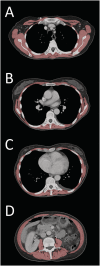
Methods: We performed a secondary analysis of prospectively collected clinical (overall survival, hospital readmission, time to unplanned hospital readmission or death, and readmission or death within 90 days) and patient-reported outcomes (physical and psychological symptom burden captured as Edmonton Symptom Assessment Scale and Patient Health Questionnaire) of patients with advanced cancer who experienced an unplanned admission to Massachusetts General Hospital from 2014 to 2016. First, we assessed the correlation of skeletal muscle cross-sectional area, SMRA, SMI, and SMG at one or more of the following thoracic (T) or lumbar (L) vertebral levels: T5, T8, T10, and L3 on CT scans obtained ≤50 days before index assessment. Second, we aggregated measurements across all available vertebral levels using percentile-based averaging (PBA) to create the average percentile. Third, we constructed one regression model adjusted for age, sex, sociodemographic factors, cancer type, body mass index, and intravenous contrast for each combination of (i) vertebral level and average percentile, (ii) muscle metrics (SMRA, SMI, & SMG), and (iii) clinical and patient-reported outcomes. Fourth, we compared the performance of vertebral levels and muscle metrics by ranking otherwise identical models by concordance statistic, number of included patients, coefficient of determination, and significance of muscle metric.
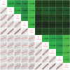
Results: We included 846 patients (mean age: 63.5 ± 12.9 years, 50.5% males) with advanced cancer [predominantly gastrointestinal (32.9%) or lung (18.9%)]. The correlation of muscle measurements between vertebral levels ranged from 0.71 to 0.84 for SMRA and 0.67 to 0.81 for SMI. The correlation of individual levels with the average percentile was 0.90-0.93 for SMRA and 0.86-0.92 for SMI. The intrapatient correlation of SMRA with SMI was 0.21-0.40. PBA allowed for inclusion of 8-47% more patients than any single-level analysis. PBA outperformed single-level analyses across all comparisons with average ranks 2.6, 2.9, and 1.6 for concordance statistic, coefficient of determination, and significance (range 1-5, μ = 3), respectively. On average, SMG outperformed SMRA and SMI across outcomes and vertebral levels: the average rank of SMG was 1.4, 1.4, and 1.4 for concordance statistic, coefficient of determination, and significance (range 1-3, μ = 2), respectively.
Conclusions: Multivertebral level skeletal muscle analyses using PBA and SMG independently and additively outperform analyses using individual levels and SMRA or SMI.
Keywords: BMI; Body composition analysis; Patient-reported outcomes; Sarcopenia; Skeletal muscle; Skeletal muscle gauge; Survival.
© 2021 The Authors. Journal of Cachexia, Sarcopenia and Muscle published by John Wiley & Sons Ltd on behalf of Society on Sarcopenia, Cachexia and Wasting Disorders.
Conflict of interest statement
F.J.F. was supported by the American Roentgen Ray Society Scholarship for this study and has a related patent pending. E.J.R., MD has served as a consultant for Mitobridge Inc., Asahi Kasei Pharmaceuticals, DRG Consulting, Napo Pharmaceuticals, American Imaging Management, Immuneering Corporation, and Prime Oncology. Additionally, he has served on recent advisory boards for Heron Pharmaceuticals, Vector Oncology, and Helsinn Pharmaceuticals. He has also served as a member on data safety monitoring boards for Oragenics, Inc, Galera Pharmaceuticals, and Enzychem Lifesciences Pharmaceutical Company. The other authors do not report relevant conflicts of interest.
Figures


References
-
- Martin L, Birdsell L, MacDonald N, Reiman T, Clandinin MT, McCargar LJ, et al. Cancer cachexia in the age of obesity: skeletal muscle depletion is a powerful prognostic factor, independent of body mass index. J Clin Oncol 2013;31:1539–1547. - PubMed
-
- Mourtzakis M, Prado CMM, Lieffers JR, Reiman T, McCargar LJ, Baracos VE. A practical and precise approach to quantification of body composition in cancer patients using computed tomography images acquired during routine care. Appl Physiol Nutr Metab 2008;33:997–1006. - PubMed
-
- Troschel AS, Troschel FM, Best TD, Gaissert HA, Torriani M, Muniappan A, et al. Computed tomography–based body composition analysis and its role in lung cancer care. J Thorac Imaging 2020;35:91–100. - PubMed
-
- Chen L‐K, Liu L‐K, Woo J, Assantachai P, Auyeung T‐W, Bahyah KS, et al. Sarcopenia in Asia: consensus report of the Asian Working Group for Sarcopenia. J Am Med Dir Assoc 2014;15:95–101. - PubMed
MeSH terms
LinkOut - more resources
Full Text Sources
Research Materials

