. 2022 Jan 26;96(2):e0090921.
doi: 10.1128/JVI.00909-21. Epub 2021 Nov 3.

Characterization of the Interaction Domains between the Phosphoprotein and the Nucleoprotein of Human Metapneumovirus

Affiliations

Characterization of the Interaction Domains between the Phosphoprotein and the Nucleoprotein of Human Metapneumovirus

Hortense Decool et al. J Virol. .

Abstract

Human metapneumovirus (HMPV) causes severe respiratory diseases in young children. The HMPV RNA genome is encapsidated by the viral nucleoprotein (N), forming an RNA-N complex (NNuc), which serves as the template for genome replication and mRNA transcription by the RNA-dependent RNA polymerase (RdRp). The RdRp is formed by the association of the large polymerase subunit (L), which has RNA polymerase, capping, and methyltransferase activities, and the tetrameric phosphoprotein (P). P plays a central role in the RdRp complex by binding to NNuc and L, allowing the attachment of the L polymerase to the NNuc template. During infection these proteins concentrate in cytoplasmic inclusion bodies (IBs) where viral RNA synthesis occurs. By analogy to the closely related pneumovirus respiratory syncytial virus (RSV), it is likely that the formation of IBs depends on the interaction between HMPV P and NNuc, which has not been demonstrated yet. Here, we finely characterized the binding P-NNuc interaction domains by using recombinant proteins, combined with a functional assay for the polymerase complex activity, and the study of the recruitment of these proteins to IBs by immunofluorescence. We show that the last 6 C-terminal residues of HMPV P are necessary and sufficient for binding to NNuc and that P binds to the N-terminal domain of N (NNTD), and we identified conserved N residues critical for the interaction. Our results allowed us to propose a structural model for the HMPV P-NNuc interaction. IMPORTANCE Human metapneumovirus (HMPV) is a leading cause of severe respiratory infections in children but also affects human populations of all ages worldwide. Currently, no vaccine or efficient antiviral treatments are available for this pneumovirus. A better understanding of the molecular mechanisms involved in viral replication could help the design or discovery of specific antiviral compounds. In this work, we have investigated the interaction between two major viral proteins involved in HMPV RNA synthesis, the N and P proteins. We finely characterized their domains of interaction and identified a pocket on the surface of the N protein, a potential target of choice for the design of compounds interfering with N-P complexes and inhibiting viral replication.

Keywords: HMPV; nucleoprotein; phosphoprotein; protein-protein interaction; structural modeling.


Figures
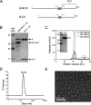
FIG 1
FIG 1
Purification of HMPV N-RNA rings using GST-PCT. (A) Schematic representation of HMPV and RSV P proteins. The oligomerization domain is represented by a rectangle and disordered N- and C-terminal domains (PNT and PCT, respectively) by lines. PCT is indicated. Residues flanking the structural domains are indicated by numbers. (B) GST-PCT and N proteins were coexpressed in E. coli, followed by purification using the GST tag. MW, molecular weight. (Left) The product of purification was analyzed by SDS-PAGE and Coomassie blue staining. (Right) The sample was then incubated in the presence of thrombin in order to cleave GST (remaining on beads) and isolate PCT-N in the supernatant. (C) Gel filtration profile of purified PCT-N complex. The curves corresponding to OD spectra at 220 nm, 260 nm, and 280 nm are presented. P1 and P2 indicate the two peaks detected. The fractions corresponding to P1 were pooled, and the sample was analyzed by SDS-PAGE colored with Coomassie blue. (D) Dynamic light scattering (DLS) analysis of the purified N protein, showing a homogenous peak at 18 nm, corresponding to N oligomers. (E) Image of purified HMPV N-RNA rings as observed by negative-stain electron microscopy. Bar, 100 nm.
FIG 2
FIG 2
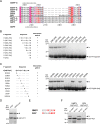
Identification of the residues of P involved in N binding. (A) Sequence alignment of Pneumoviridae P C terminus (PCT), from the Orthopneumovirus and Metapneumovirus genera: hRSV-A/-B (human RSV strains A and B, UniProt entries PHOSP_HRSVA and PHOSP_HRSVB, respectively), oRSV (ovine RSV, UniProt entry PHOSP_ORSVW), bRSV (bovine RSV, UniProt entry PHOSP_BRSVA), mPV (murine pneumonia virus, Uniprot entry PHOSP_MPV15), aMPV-A/-C (avian metapneumovirus type A and C, UniProt entries Q65032 and PHOSP_AMPV1, respectively), and hMPV (human metapneumovirus strain CAN97-83, UniProt entry PHOSP_HMPVC). Sequence numbering and secondary structures are indicated at the top for hRSV and at the bottom for hMPV. (B) GST-P fragments and C/GST-P[285-294] (WT or mutants) (sequences indicated on the left) were coexpressed with N in bacteria, followed by purification using the GST tag. The products of purification were analyzed by SDS-PAGE and Coomassie blue staining (right). (D) GST-PCT or GST-PCTALAM corresponding to the double substitution of residues Q291 and I293 of P by alanine were coexpressed with N. The products of purification by GST were analyzed by SDS-PAGE and Coomassie blue staining. (E) Sequence alignment of the last 9 C-terminal residues of HMPV and RSV P proteins. The residues critical for the interaction with the N protein are indicated in red boldface. F/HMPV and RSV GST-PCT (P residues 200 to 294 for HMPV and P residues 161 to 241 for RSV) constructs were coexpressed with either HMPV or RSV N proteins. Copurification of N proteins by GST-PCT fragment was analyzed by SDS-PAGE and Coomassie blue staining.
FIG 3
FIG 3
Search for the PCT binding site on HMPV N. (A) Amino acid sequence alignments between the N-terminal domains of N (residues 1 to 252/253) proteins of HMPV (strain CAN 97-83) and human RSV (HRSV, strain Long VR-26). Invariant residues are highlighted in white font on a red background. The secondary structure elements observed in the crystal structure of HMPV N protein (11) are indicated above the sequence. Green asterisks below the sequence indicate the residues constituting the P binding pocket of RSV N. UniProt accession codes were NCAP_HRSVA (human RSV) and A1DZS3_9MONO (human MPV). Sequences were aligned with Clustal W and treated with ESPript 3. (B) Side view (left) and bottom view (right) of the HMPV N-RNA rings (PDB accession number 5FVC) showing one N monomer colored according to domains: N arm in blue, C arm in green, and N- and C-terminal domains (NNTD and NCTD, respectively) in yellow and red. The residues identified by analogy with RSV as involved in P binding on NNTD are indicated by black spheres.
FIG 4
FIG 4
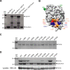
Identification of the residues of NNTD involved in P binding. (A) GST-P or GST-PΔM294 and NNTD-6xHis proteins were coexpressed in E. coli, followed by purification using the 6×His tag. GST-P or GST-PΔM294 alone was purified using the GST tag. The products of purification were analyzed by SDS-PAGE and Coomassie blue staining. (B) Surface representation of HMPV NNTD (from Gly30 to Gly 253, indicated in green) showing the potential P binding pocket, with acidic amino acids colored in red, basic residues in blue, and hydrophobic residues in orange. (C) The impact of N mutations on the expression and solubility of the protein was assessed by expressing 6xHis-N protein (WT or mutants) in E. coli, followed by purification and SDS-PAGE analysis. (D) GST-PCT and mutant N proteins were coexpressed in E. coli, followed by purification using the GST tag. The products of purification were analyzed by SDS-PAGE and Coomassie blue staining (upper), and the expression of N proteins in bacteria was validated by Western blotting (lower).
FIG 5
FIG 5
Residues involved in P-N-RNA ring interaction are critical for polymerase activity. (A) HMPV polymerase activity in the presence of P and N mutants. BSRT7/5 cells were transfected with plasmids encoding the P (WT or mutants), N (WT, or mutants), L, and M2-1 proteins and the pGaussia/Firefly minigenome, together with pCMV-βGal for transfection standardization. Viral RNA synthesis was quantified by measuring the Gaussia luciferase activity after cell lysis 24 h after transfection. Each luciferase minigenome activity value was normalized based on β-galactosidase expression and is the average from three independent experiments performed in triplicate. Error bars represent standard deviations calculated based on two independent experiments made in quadruplicate. (B) Western blot showing the expression of P and N proteins (WT and mutants) in BSRT7/5 cells. (C and D) HMPV polymerase activity in the presence of pN–pGFP-N (at a ratio of 2:1) or P-BFP. Error bars represent standard deviations calculated based on two independent experiments made in triplicate. The statistical significance of differences was calculated using Student's t test. ***, P < 0.001; ****, P < 0.001; ns, not significant.
FIG 6
FIG 6
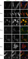
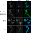
Impact of P mutations on the formation of HMPV cytoplasmic IBs. BSRT7/5 cells transfected with plasmids encoding P (WT or mutants) and N + GFP-N (ratio, 2:1) proteins were fixed 24 h posttransfection and labeled with anti-P antibody, and the distribution of viral proteins was observed by fluorescence microscopy. Nuclei were stained with Hoechst 33342. Scale bars, 10 μm.
FIG 7
FIG 7
Impact of N mutations on the formation of HMPV cytoplasmic IBs. BSRT7/5 cells transfected with plasmids encoding P-BFP and N (WT or mutants) proteins were fixed 24 h posttransfection and labeled with anti-N antibody, and the distribution of viral proteins was observed by fluorescence microscopy. Scale bars, 10 μm.
FIG 8
FIG 8
Ten best-scoring structures of clusters 1 (A) and 2 (B) from HADDOCK refinement of HMPV P3 (residues L292-I293-M294 of P)/NNTD complex superimposed with RSV P2/NNTD. NNTD is colored in beige (HMPV) or yellow (RSV), HMPV P3 peptide in cyan (HMPV), and RSV P2 peptide (corresponding to residues D240-F241 of P) in pink. Side chains of residues in contact with the P peptide in HADDOCK models are also shown, along with their RSV equivalent.
FIG 9
FIG 9
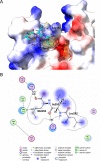
Model of HMPV P3/NNTD complex from HADDOCK refinement. (A) The NNTD protein is colored by electrostatic surface, and the P3 peptide (residues L292-I293-M294 of P) is shown as sticks. The residues of NNTD critical for the interaction with P are indicated, with residues for mutation to alanine that abrogate P binding labeled in boldface. Yellow surface shows InDeep hydrophobic channel prediction where hydrophobic contacts are expected to occur at the NNTD surface. (B) Interaction diagram between P3 peptide (Leu292-Ile293-Met294) and NNTD.
FIG 10
FIG 10
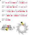
Conservation of pneumovirus N and P residues. (A) Sequence logo of the last 9 C-terminal residues of P of Metapneumovirus (left) and Orthopneumovirus (right) sequences. P sequences were collected from Interpro entry IPR003487 (464 unique sequences), and logos were created from nonredundant peptide sequences of the last 9 amino acids. (B) Sequence logo of the residues forming the P-binding site on N of the Metapneumovirus (left) and Orthopneumovirus (right) genera. N sequences were collected from Interpro entry IPR004930 (359 unique sequences) and aligned. Classification as ortho- and metapneumovirus was based on UniProt taxonomy (taxon identifiers 1868215 for OrthoPV and 162387 for MetaPV). Residue numbers are shown at the bottom, and gray markers indicate residues identified as essential for P-N interaction. Logos were generated with WebLogo (29).

References

    1. Pneumonia Etiology Research for Child Health Study Group. 2019. Causes of severe pneumonia requiring hospital admission in children without HIV infection from Africa and Asia: the PERCH multi-country case-control study. Lancet 394:757–779. 10.1016/S0140-6736(19)30721-4. - DOI - PMC - PubMed
    1. Schildgen V, van den Hoogen B, Fouchier R, Tripp RA, Alvarez R, Manoha C, Williams J, Schildgen O. 2011. Human Metapneumovirus: lessons learned over the first decade. Clin Microbiol Rev 24:734–754. 10.1128/CMR.00015-11. - DOI - PMC - PubMed
    1. van den Hoogen BG, de Jong JC, Groen J, Kuiken T, de Groot R, Fouchier RA, Osterhaus AD. 2001. A newly discovered human pneumovirus isolated from young children with respiratory tract disease. Nat Med 7:719–724. 10.1038/89098. - DOI - PMC - PubMed
    1. Afonso CL, Amarasinghe GK, Bányai K, Bào Y, Basler CF, Bavari S, Bejerman N, Blasdell KR, Briand F-X, Briese T, Bukreyev A, Calisher CH, Chandran K, Chéng J, Clawson AN, Collins PL, Dietzgen RG, Dolnik O, Domier LL, Dürrwald R, Dye JM, Easton AJ, Ebihara H, Farkas SL, Freitas-Astúa J, Formenty P, Fouchier RAM, Fù Y, Ghedin E, Goodin MM, Hewson R, Horie M, Hyndman TH, Jiāng D, Kitajima EW, Kobinger GP, Kondo H, Kurath G, Lamb RA, Lenardon S, Leroy EM, Li C-X, Lin X-D, Liú L, Longdon B, Marton S, Maisner A, Mühlberger E, Netesov SV, Nowotny N, et al.. 2016. Taxonomy of the order Mononegavirales: update 2016. Arch Virol 161:2351–2360. 10.1007/s00705-016-2880-1. - DOI - PMC - PubMed
    1. van den Hoogen BG, Bestebroer TM, Osterhaus AD, Fouchier RA. 2002. Analysis of the genomic sequence of a human metapneumovirus. Virology 295:119–132. 10.1006/viro.2001.1355. - DOI - PubMed

Publication types

MeSH terms