Case Reports
. 2022 Jan;104(1):e21-e24.
doi: 10.1308/rcsann.2021.0099. Epub 2021 Nov 3.

Core needle biopsy causing a pseudoaneurysm in the breast

Affiliations
Case Reports

Core needle biopsy causing a pseudoaneurysm in the breast

B Swain et al. Ann R Coll Surg Engl. 2022 Jan.

Abstract

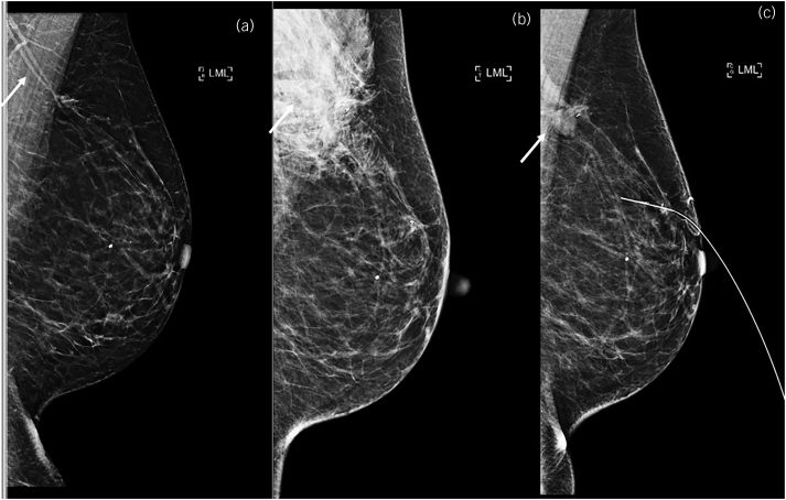
Core needle biopsy (CNB) is the first-choice method of sampling suspicious, focal breast lesions for histological analysis. Here we present the case of a 65-year-old woman who was recalled for evaluation of the left breast following a screening mammogram. An ultrasound-guided biopsy was performed using a disposable core biopsy needle and 3 weeks later a magnetic resonance imaging scan showed a distended vessel with adjacent sac measuring 17 × 15mm2. A Doppler ultrasound scan confirmed pseudoaneurysm. A review of the literature was made on breast pseudoaneurysm following CNB, and over the past 20 years there were few other reports. Pseudoaneurysms in the breast are a rare but serious complication of CNBs. They may spontaneously thrombose, but often require intervention, so it is essential that clinicians are aware of the risk.

Keywords: Breast; Core needle biopsy; Pseudoaneurysm.


Figures

Figure 1
Figure 1
Craniocaudal screening mammogram showing lesions A and B
Figure 2
Figure 2
Mediolateral oblique screening mammogram again showing lesions A and B
Figure 3
Figure 3
Comparative lateromedial (LM) mammograms. (a) Synthetic LM mammogram (C-view). The arrow demonstrates proximity of vessels to the tumour. (b) Post-biopsy LM mammogram. The arrow demonstrates the post-biopsy haematoma. Note metallic clip placement into the tumours A and B. (c) Preoperative LM mammogram showing wire placed into lesion A. The arrow shows the smooth contour of the pseudoaneurysm arising from the vessel marked in (a) which is now distended.
Figure 4
Figure 4
Axial T1 post-contrast minimum intensity projection magnetic resonance image with an arrow showing the distended feeding vessel and pseudoaneurysm in the upper outer quadrant of the left breast
Figure 5
Figure 5
Sagittal T1 post-contrast magnetic resonance image with an arrow showing the pseudoaneurysm and feeding vessel
Figure 6
Figure 6
Longitudinal ultrasound image showing the well-defined and anechoic pseudoaneurysm
Figure 7
Figure 7
Transverse ultrasound image showing the well-defined and anechoic pseudoaneurysm
Figure 8
Figure 8
Transverse Doppler ultrasound image showing a ‘yin–yang’ sign indicating bidirectional flow because of the swirling of blood, which is a radiological sign described in pseudoaneurysm. The ill-defined hypoechoic shadowing of the malignant mass is noted just medial to the pseudoaneurysm.

References

    1. NHS Breast Cancer Screening Program (NHSBSP). Clinical Guidance for Breast Cancer Screening Assessment. NHSBSP Publication No 49. PHE publications gateway number 2016426. 2016 Nov.
    1. El Khoury M, Mesurolle B, Kao Eet al. . Spontaneous thrombosis of pseudoaneurysm of the breast related to core biopsy. Am J Roentgenol 2007; 189: W309–W311.10.2214/AJR.05.1647 - DOI - PubMed
    1. Mahoney MC, Ingram AD. Breast emergencies: types, imaging features, and management. AJR Am J Roentgenol 2014; 202: W390–W399.10.2214/AJR.13.11758 - DOI - PubMed
    1. Adler K, Samreen N, Glazebrook KN, Bhatt AA. Imaging features and treatment options for breast pseudoaneurysms after biopsy: A case-based pictorial review. J Ultrasound Med 2020; 39: 181–190.10.1002/jum.15082 - DOI - PubMed
    1. Russell T, Creagh-Barry M. Breast pseudoaneurysm arising from core needle biopsy should be left well alone. BMJ Case Rep 2017; 2017: bcr2017221546.10.1136/bcr-2017-221546 - DOI - PMC - PubMed

Publication types