Review
. 2021 Dec;78(24):7925-7942.
doi: 10.1007/s00018-021-03984-7. Epub 2021 Nov 3.

Nucleotide excision repair leaves a mark on chromatin: DNA damage detection in nucleosomes

Affiliations
Review

Nucleotide excision repair leaves a mark on chromatin: DNA damage detection in nucleosomes

Katja Apelt et al. Cell Mol Life Sci. 2021 Dec.

Abstract

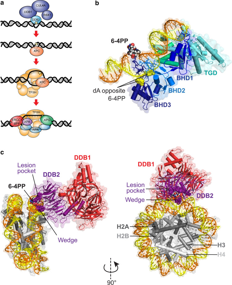
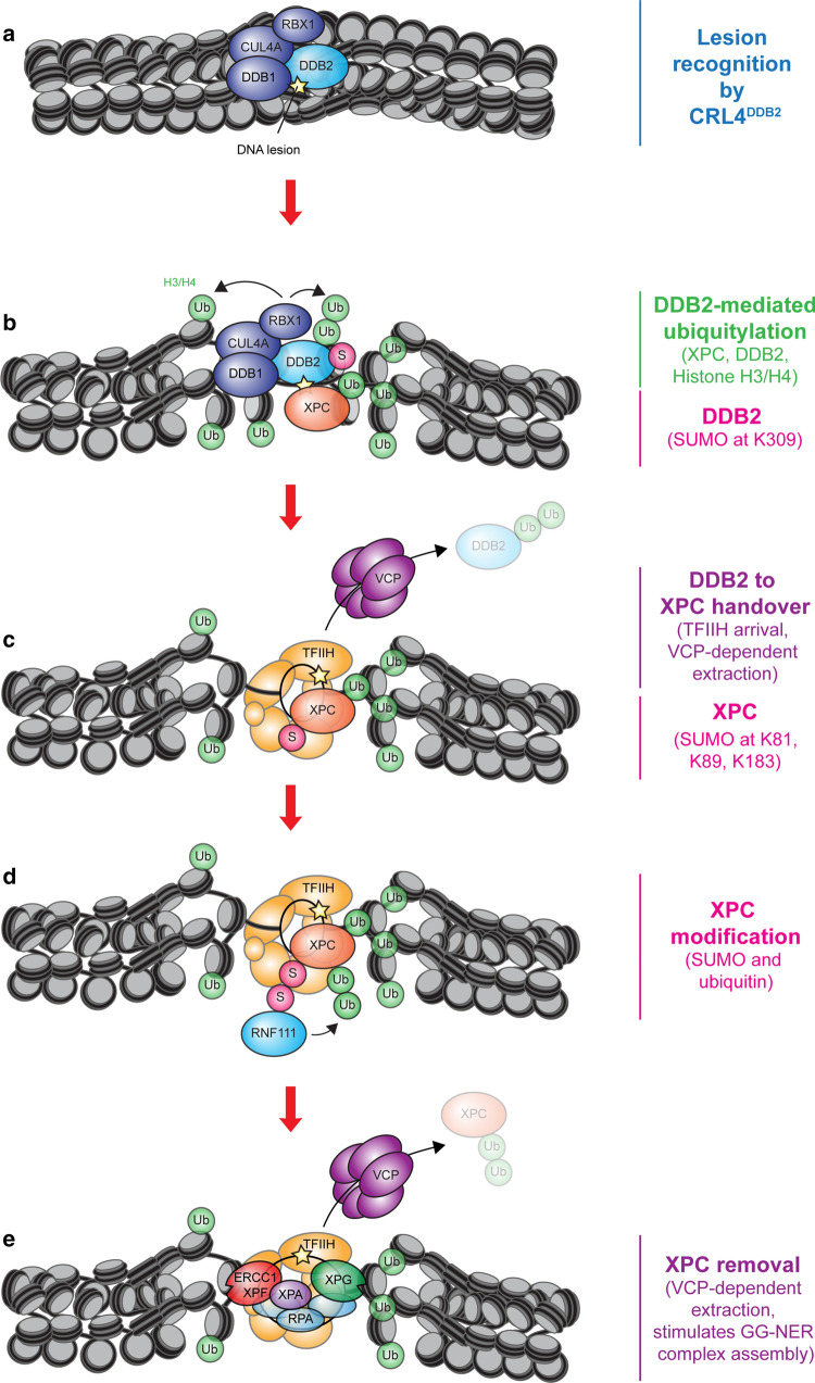
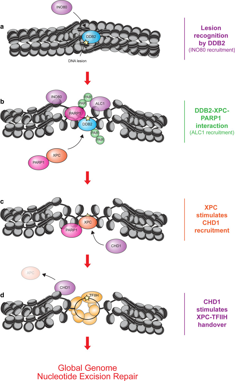
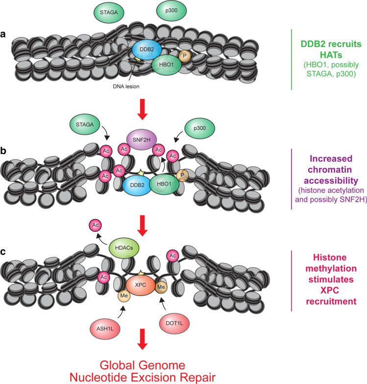
Global genome nucleotide excision repair (GG-NER) eliminates a broad spectrum of DNA lesions from genomic DNA. Genomic DNA is tightly wrapped around histones creating a barrier for DNA repair proteins to access DNA lesions buried in nucleosomal DNA. The DNA-damage sensors XPC and DDB2 recognize DNA lesions in nucleosomal DNA and initiate repair. The emerging view is that a tight interplay between XPC and DDB2 is regulated by post-translational modifications on the damage sensors themselves as well as on chromatin containing DNA lesions. The choreography between XPC and DDB2, their interconnection with post-translational modifications such as ubiquitylation, SUMOylation, methylation, poly(ADP-ribos)ylation, acetylation, and the functional links with chromatin remodelling activities regulate not only the initial recognition of DNA lesions in nucleosomes, but also the downstream recruitment and necessary displacement of GG-NER factors as repair progresses. In this review, we highlight how nucleotide excision repair leaves a mark on chromatin to enable DNA damage detection in nucleosomes.

Keywords: Chromatin; DDB2; Nucleotide excision repair; PTM; Post-translational modification; XPC.


Conflict of interest statement

The authors declare that they have no conflict of interest.

Figures

Fig. 1
Fig. 1
DNA damage-recognition factors initiate GG-NER. a Model of GG-NER initiation by the CRL4DDB2 complex (consisting of DDB2-DDB1-CUL4A-RBX1) and the XPC complex (consisting of XPC, RAD23B, CETN2), which is followed by the recruitment of the TFIIH complex, XPA, RPA and the endonucleases XPG and ERCC1-XPF. b Structure of yeast Rad4/XPC bound to a 6-4PP lesion. The lesion is displaced from the helix stack using the hairpins of the BHD2 and BHD3 domains. The BHD2/3 domains form a tight binding pocket for the dA residues in the non-damaged strand, but do not contact the lesion directly. The BHD1-TGD domains of Rad4/XPC bind in a damage and sequence non-specific manner and anchor the protein on DNA during the lesion search process. Figure generated using PDB 6CFI with PyMol. c Structure of UV-DDB (consisting of DDB2 and DDB1) bound to a 6-4PP in a nucleosome. The DDB2 protein binds to the nucleosome at a 60° angle and pushes the 6-4PP into a lesion-binding pocket using wedge residues (F334, Q335 and H336). Figure generated using PDB 6R8Y with PyMol
Fig. 2
Fig. 2
The role of ubiquitylation and SUMOylation in GG-NER. a DDB2 is part of the CRL4DDB2 ubiquitin ligase complex together with DDB1, CUL4A and RBX1 that binds to photolesions. b The CRL4DDB2 ligase ubiquitylates H3 and H4 leading to chromatin decompaction through an unknown mechanism, which stimulates XPC recruitment. DDB2 also ubiquitylates XPC, which increases its affinity for DNA lesions. DDB2 becomes SUMOylated at K309, which stimulates XPC recruitment and promotes CPD repair. c DDB2 must dissociate to allow stable XPC binding to the DNA lesion. The displacement of DDB2 is stimulated by the recruitment of TFIIH by XPC. The ubiquitin-selective segregase VCP/p97 further stimulates the dissociation of DDB2 through extraction of ubiquitylated DDB2 from chromatin. XPC is SUMOylated at K81, K89 and K183, which was suggested to stimulate the handover between XPC and DDB2. d The SUMO-dependent E3 ubiquitin ligase RNF111 recognizes and ubiquitylates the SUMOylated form of XPC. e The ubiquitylated form of XPC may also be extracted from chromatin by VCP/p97 to enable efficient recruitment of the endonuclease XPG. This is important because XPG and XPC cannot coexist in the same NER complex
Fig. 3
Fig. 3
The role of chromatin remodelers and PARylation in GG-NER. a Lesion recognition by DDB2 may recruit the ATP-dependent chromatin remodeler INO80. While INO80 was shown to be recruited by DDB1, we speculate that this is also dependent on DDB2. b DDB2 interacts with PARP1 and stimulates its catalytic activity. Note that PARP1 binds to photolesions independently of DDB2 or XPC. PARP1 modifies itself and DDB2 with PAR chains. The PAR-binding chromatin remodeler ALC1 is recruited and stimulates GG-NER. PARP1 also interacts with XPC already in the absence of DNA damage and facilitates its recruitment to photolesions, particularly at low damage load. c XPC recruits the chromatin remodeler CHD1. d CHD1 facilitates the displacement of XPC to stimulate TFIIH recruitment
Fig. 4
Fig. 4
A model of the role of acetylation, deacetylation and methylation in GG-NER. a UV-induced lesions in chromatin are recognized by DDB2 resulting in the recruitment of three histone acetyltransferases (STAGA, p300 and phosphorylated HBO1). b These enzymes catalyse histone acetylation leading to increased chromatin accessibility. Phosphorylated HBO1 recruits the chromatin remodeler SNF2H. c Efficient recruitment of XPC also requires histone deacetylation, which is facilitated by DDB2 through the proteolytic degradation of p300 and HBO1, as well as through the recruitment of histone deacetylases (HDAC1-4). Finally, DDB2 also recruits the methyltransferase ASH1L and possibly DOT1L, which methylate H3K4 and H3K79, respectively. XPC preferentially associates with nucleosomes containing methylated histones

References

    1. Gates KS. An overview of chemical processes that damage cellular DNA: spontaneous hydrolysis, alkylation, and reactions with radicals. Chem Res Toxicol. 2009;22:1747–1760. - PMC - PubMed
    1. Hoeijmakers JH. DNA damage, aging, and cancer. N Engl J Med. 2009;361:1475–1485. - PubMed
    1. Ciccia A, Elledge SJ. The DNA damage response: making it safe to play with knives. Mol Cell. 2010;40:179–204. - PMC - PubMed
    1. Marteijn JA, Lans H, Vermeulen W, Hoeijmakers JH. Understanding nucleotide excision repair and its roles in cancer and ageing. Nat Rev Mol Cell Biol. 2014;15:465–481. - PubMed
    1. van den Heuvel D, van der Weegen Y, Boer DEC, Ogi T, Luijsterburg MS. Transcription-coupled DNA Repair: from mechanism to human disorder. Trends Cell Biol. 2021 doi: 10.1016/j.tcb.2021.02.007. - DOI - PubMed

LinkOut - more resources