. 2021 Nov 3;12(1):158.
doi: 10.1186/s13244-021-01097-0.

Limitations of cardiothoracic ratio derived from chest radiographs to predict real heart size: comparison with magnetic resonance imaging

Affiliations

Limitations of cardiothoracic ratio derived from chest radiographs to predict real heart size: comparison with magnetic resonance imaging

Paulius Simkus et al. Insights Imaging. .

Abstract

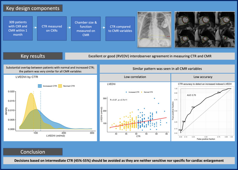
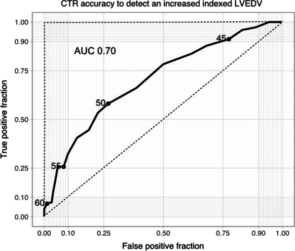
Background: Cardiothoracic ratio (CTR) in chest radiographs is still widely used to estimate cardiac size despite the advent of newer imaging techniques. We hypothesise that a universal CTR cut-off value of 50% is a poor indicator of cardiac enlargement. Our aim was to compare CTR with volumetric and functional parameters derived from cardiac magnetic resonance imaging (MRI).

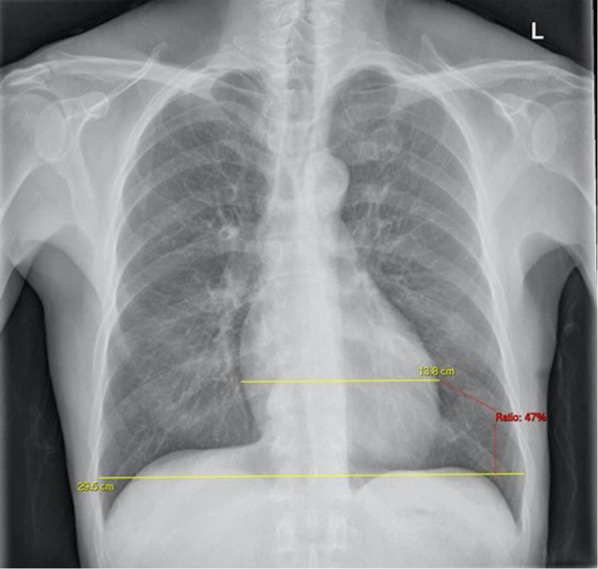
Methods: 309 patients with a chest radiograph and cardiac MRI acquired within a month were reviewed to assess how CTR correlates with multiple cardiac MRI variables: bi-ventricular EDV (absolute and indexed to body surface area), EF, indexed total heart volume and bi-atrial areas. In addition, we have also determined CTR accuracy by creating multiple ROC curves with the described variables.

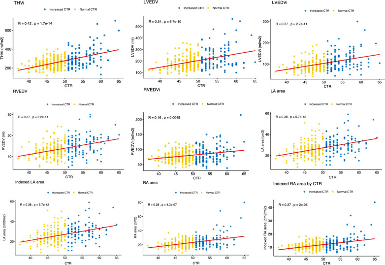
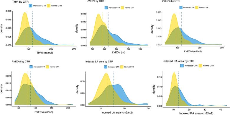
Results: All cardiac MRI variables correlate weakly but statistically significantly with CTR. This weak correlation is explained by a substantial overlap of cardiac MRI parameters in patients with normal and increased CTR. For all variables, CTR was only mildly to moderately better than a chance to discriminate cardiac enlargement (AUC 0.6-0.7). Large CTR values (> 55%) are specific but not sensitive, while low CTR values (< 45%) are sensitive but not specific. Values in between are not sensitive nor specific.

Conclusions: CTR correlates weakly with true chamber size assessed by gold standard cardiac MRI and has a weak discriminatory power. Thus, clinical decisions based on intermediate CTRs (45-55%) should be avoided. Large CTRs (> 55%) are likely indicative of true heart chamber enlargement. Low CTRs (< 45%) are likely indicative of normal heart size.

Keywords: Cardiac imaging; Cardiac magnetic resonance imaging; Cardiothoracic ratio; Chest radiograph.


Conflict of interest statement

The authors declare that they have no competing interests.

Figures

Fig. 1
Fig. 1
Key components of study design and results
Fig. 2
Fig. 2
The cardiothoracic ratio on PA chest radiograph. The maximum transverse cardiac diameter is divided by the maximum transverse diameter of the thorax and multiplied by 100
Fig. 3
Fig. 3
Scatterplots including correlation lines between CTR and cardiac volumes and areas. Yellow dots represent patients with CTR < 50%, blue dots patients with CTR > 50%. Although there is a weak trend for patients with larger CTR to have larger ventricular volumes and atrial areas, the majority of measured cardiac MRI parameters are noted in both individuals with normal and increased CTR. THVi total heart indexed volume, LVEDV left ventricular end-diastolic volume, LVEDVi left ventricular indexed end-diastolic volume, RVEDV right ventricular end-diastolic volume, RVEDVi right ventricular indexed end-diastolic volume, LA left atrium, CTR cardiothoracic ratio
Fig. 4
Fig. 4
Density plots representing distributions of cardiac MRI parameters in patients with normal and increased CTR. Density plots are a variation of histograms that allow to observe the distribution of a variable in a dataset in a continuous fashion. They are instrumental in demonstrating the extensive overlap of cardiac MRI derived values between patients with normal and increased CTR. Dashed lines represent the means for each distribution. Note that mean values are larger in patients with CTR > 50%. This explains the described positive but weak correlations. THVi total heart indexed volume, LVEDV left ventricular end-diastolic volume, LVEDVi left ventricular indexed end-diastolic volume, RVEDVi right ventricular indexed end-diastolic volume, LA left atrium, RA right atrium, CTR cardiothoracic ratio
Fig. 5
Fig. 5
ROC curve of CTR and increased LVEDV. ROC curve illustrating diagnostic accuracy of multiple CTR cut-off values to detect an increased LVEDVi. The X-axis shows true positives for each cut-off (an increased LVEDV on cardiac MRI is seen by the defined cut-off). The Y-axis shows false positives (normal LVEDV on cardiac MRI, which is characterised as increased based on a given CTR value). Higher CTR values give few false positives at the expense of many false negatives (many patients with an increased LVEDV are missed). Lower CTR values diagnose most patients with an increased LVEDV at the expense of many false-positive results. The area under the ROC curve describes the overall diagnostic power of CTR. An ideal test would have no false positives or false negatives (AUC = 1 described by the 90° dashed line). Random guessing would render 50% of true positives and 50% of false positives (AUC = 0.5) and is represented by the no-discrimination line (45° dashed diagonal line). CTR cardiothoracic ratio, LVEDVi left ventricular indexed end-diastolic volume, AUC area under the curve. Other parameters are available in Additional file 1: Fig. S1

References

    1. Danzer CS (1919) The cardiothoracic ratio: an index of cardiac enlargement. Am J Med Sci 513–521
    1. Grotenhuis HB, Zhou C, Tomlinson G, et al. Cardiothoracic ratio on chest radiograph in pediatric heart disease: How does it correlate with heart volumes at magnetic resonance imaging? Pediatr Radiol. 2015;45:1616–1623. doi: 10.1007/s00247-015-3386-9. - DOI - PubMed
    1. Puddy E, Hill C. Interpretation of the chest radiograph. Contin Educ Anaesth Crit Care Pain. 2007;7:71–75. doi: 10.1093/bjaceaccp/mkm014. - DOI
    1. Kelly B. The chest radiograph. Ulster Med J. 2012;81:143–148. - PMC - PubMed
    1. Zaman MJS, Sanders J, Crook AM, et al. Cardiothoracic ratio within the “normal” range independently predicts mortality in patients undergoing coronary angiography. Heart. 2007;93:491–494. doi: 10.1136/hrt.2006.101238. - DOI - PMC - PubMed

LinkOut - more resources