. 2022 Jan;23(1):27-35.
doi: 10.1007/s10048-021-00666-1. Epub 2021 Nov 3.

Myoclonic status epilepticus and cerebellar hypoplasia associated with a novel variant in the GRIA3 gene

Affiliations

Myoclonic status epilepticus and cerebellar hypoplasia associated with a novel variant in the GRIA3 gene

Berardo Rinaldi et al. Neurogenetics. 2022 Jan.

Erratum in

Abstract

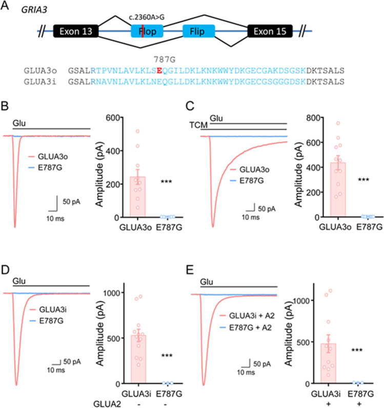
AMPA-type glutamate receptors (AMPARs) are postsynaptic ionotropic receptors which mediate fast excitatory currents. AMPARs have a heterotetrameric structure, variably composed by the four subunits GluA1-4 which are encoded by genes GRIA1-4. Increasing evidence support the role of pathogenic variants in GRIA1-4 genes as causative for syndromic intellectual disability (ID). We report an Italian pedigree where some male individuals share ID, seizures and facial dysmorphisms. The index subject was referred for severe ID, myoclonic seizures, cerebellar signs and short stature. Whole exome sequencing identified a novel variant in GRIA3, c.2360A > G, p.(Glu787Gly). The GRIA3 gene maps to chromosome Xq25 and the c.2360A > G variant was transmitted by his healthy mother. Subsequent analysis in the family showed a segregation pattern compatible with the causative role of this variant, further supported by preliminary functional insights. We provide a detailed description of the clinical evolution of the index subjects and stress the relevance of myoclonic seizures and cerebellar syndrome as cardinal features of his presentation.

Keywords: AMPARs; Cerebellar hypoplasia; GRIA3; Glutamate; Myoclonic status epilepticus.


Figures

Fig. 1
Fig. 1
Pedigree of the index family. Symptomatic individuals are shown in grey; the black arrow identifies the index subject; dashed circles identify possible female carriers
Fig. 2
Fig. 2
EEG and electromyographic polygraphy during myoclonic status epilepticus (recorded at the age of 5 years, amplitude: 400 microVolt/cm)
Fig. 3
Fig. 3
Sagittal T1-weighted cerebral MRI scan showing vermian hypoplasia
Fig. 4
Fig. 4
Facial appearance of the index subject at the age of 11 (left) and 18 (right)
Fig. 5
Fig. 5
Conservation and localization of the Glu787 residue. (a) Phylogenetic conservation among different species; (b) conservation among the primary structure of human GluA1-4 subunits; (c) graphical representation of the GluA3 subunit, published mutated residues 18, 19, 20, 22, 23, 25 are shown in blue, the Glu787 residue in red. The green residues constitute the ligand-binding domain (LBD). Figure 5a, 5b and 5c were prepared with the help of COBALT (NCBI), UniProt and Protter, respectively (please refer to Web Resources)
Fig. 6
Fig. 6
Glutamate-induced currents in wild-type GluA3 and in the E787G mutant. (A) The flip and flop of GLUA3. (B) Glutamate-induced currents in GLUA3o and GLUA3o_E787G. Left panel, sample traces. Right panel, bar graph showing the glutamate-induced current amplitudes. GLUA3o, 242.6 ± 44.5 pA, n = 10; GLUA3o_E787G, 2.9 ± 0.3 pA, n = 6. (C) Glutamate-induced currents in GLUA3o and GLUA3o_E787G in the presence of TMC. GLUA3o, 436.2 ± 56.7 pA, n = 11; GLUA3o_E787G, 2.6 ± 0.4 pA, n = 9. (D) Glutamate-induced currents in GLUA3i and GLUA3i-E787G. GLUA3i, 528.7 ± 67.7 pA, n = 12; GLUA3i_E787G, 3.6 ± 0.4, n = 3. (E) Glutamate-induced currents in GLUA3i and GLUA3i_E787G with the coexpression of GLUA2i. GLUA2/A3i, 477.1 ± 107.2 pA, n = 11; GLUA2/A3i_E787G, 7.8 ± 1.2, n = 3. Data are presented as means ± SEM. ***p < 0.001, unpaired t-test

References

    1. Beneyto M, Meador-Woodruff JH. Expression of transcripts encoding AMPA receptor subunits and associated postsynaptic proteins in the macaque brain. J Comp Neurol. 2004;468(4):530–554. doi: 10.1002/cne.10981. - DOI - PubMed
    1. Pickard L, Noel J, Henley JM, Collingridge GL, Molnar E. Developmental changes in synaptic AMPA and NMDA receptor distribution and AMPA receptor subunit composition in living hippocampal neurons. J Neurosci. 2000;20(21):7922–7931. doi: 10.1523/jneurosci.20-21-07922.2000. - DOI - PMC - PubMed
    1. Sommer B, Keinänen K, Verdoorn TA, Wisden W, Burnashev N, Herb A, et al. Flip and flop: a cell-specific functional switch in glutamate-operated channels of the CNS. Science. 1990;249(4976):1580–1585. doi: 10.1126/science.1699275. - DOI - PubMed
    1. Greger IH, Khatri L, Kong X, Ziff EB. AMPA receptor tetramerization is mediated by Q/R editing. Neuron. 2003;40(4):763–774. doi: 10.1016/S0896-6273(03)00668-8. - DOI - PubMed
    1. Petrini EM, Lu J, Cognet L, Lounis B, Ehlers MD, Choquet D. Endocytic trafficking and recycling maintain a pool of mobile surface AMPA receptors required for synaptic potentiation. Neuron. 2009;63(1):92–105. doi: 10.1016/j.neuron.2009.05.025. - DOI - PMC - PubMed

Publication types

Supplementary concepts