. 2021 Nov 2;37(5):109917.
doi: 10.1016/j.celrep.2021.109917.

Temporal dynamics of base excision/single-strand break repair protein complex assembly/disassembly are modulated by the PARP/NAD+/SIRT6 axis

Affiliations

Temporal dynamics of base excision/single-strand break repair protein complex assembly/disassembly are modulated by the PARP/NAD+/SIRT6 axis

Christopher A Koczor et al. Cell Rep. .

Abstract

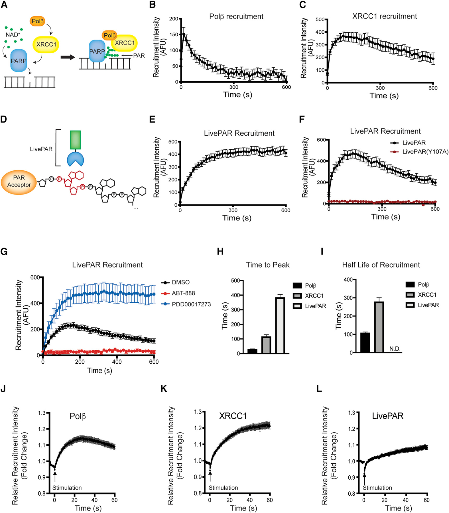
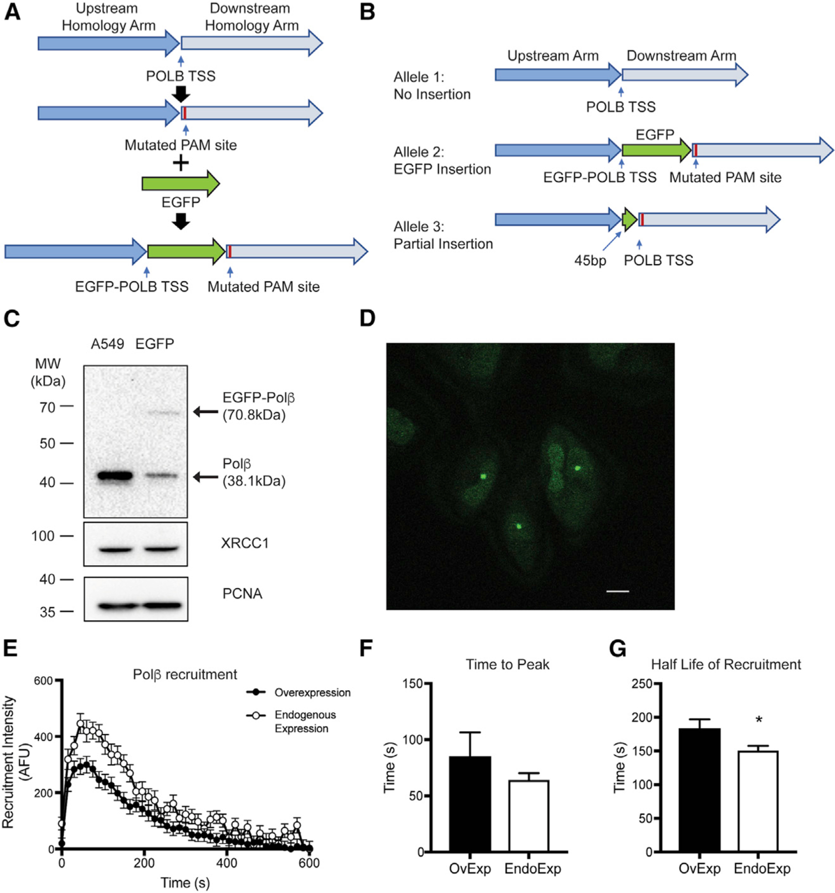
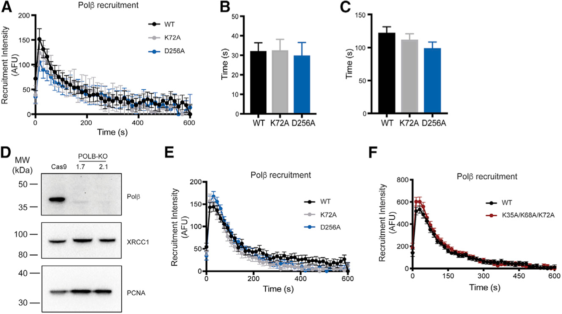
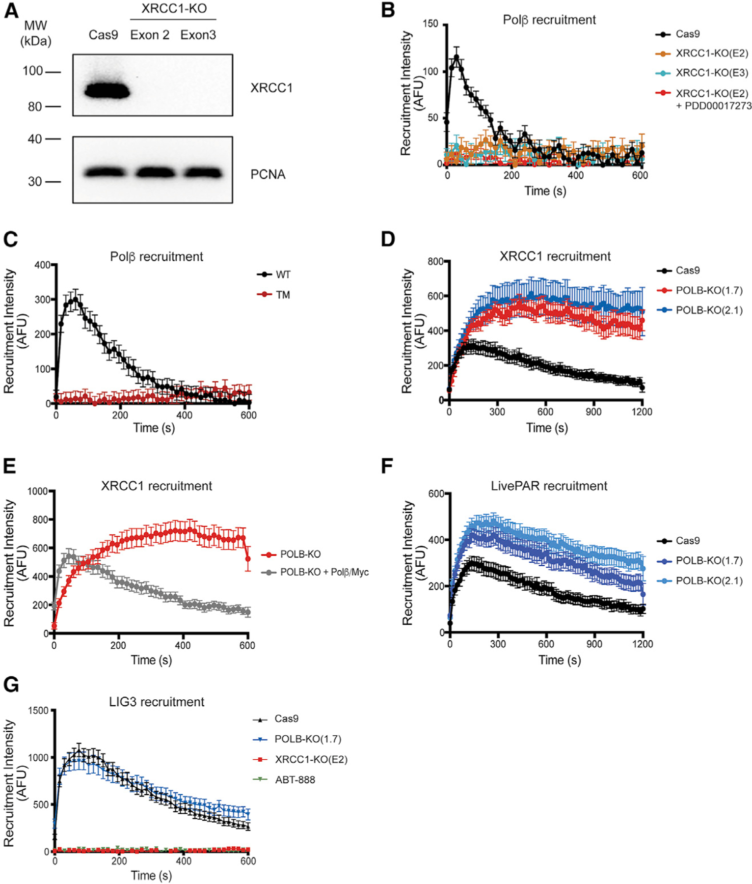
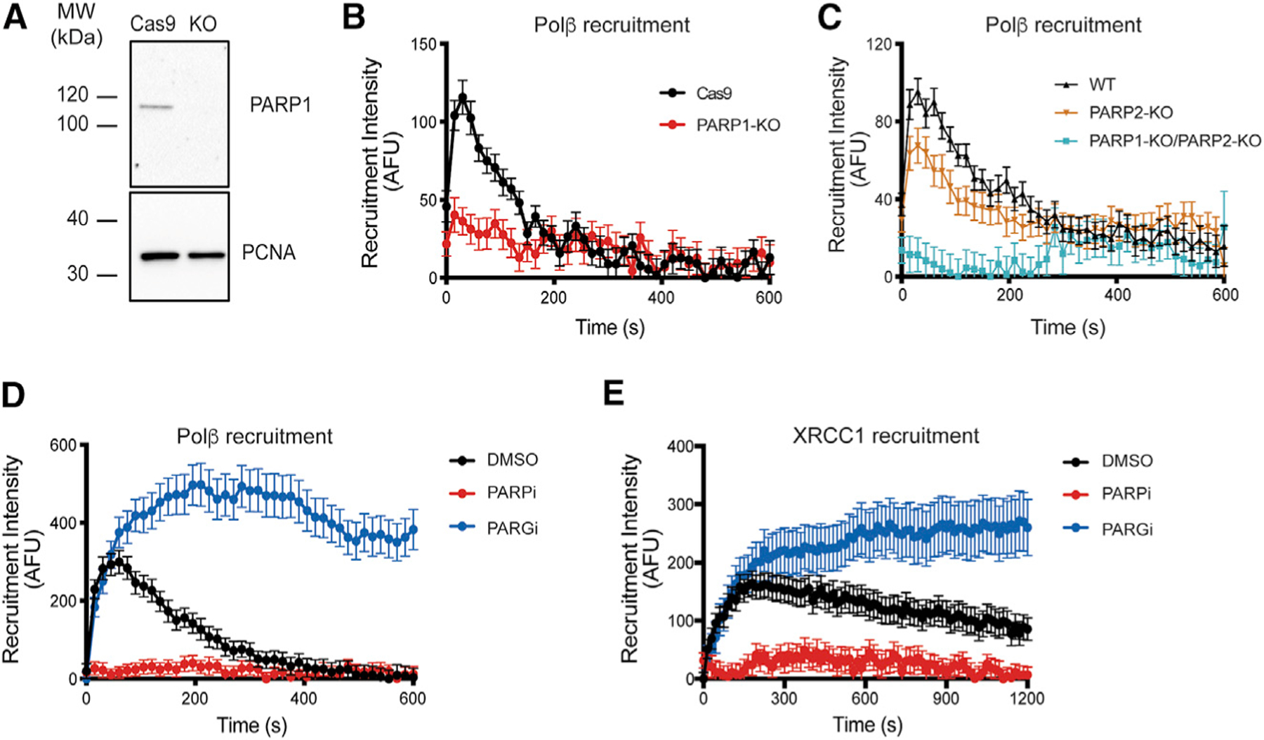
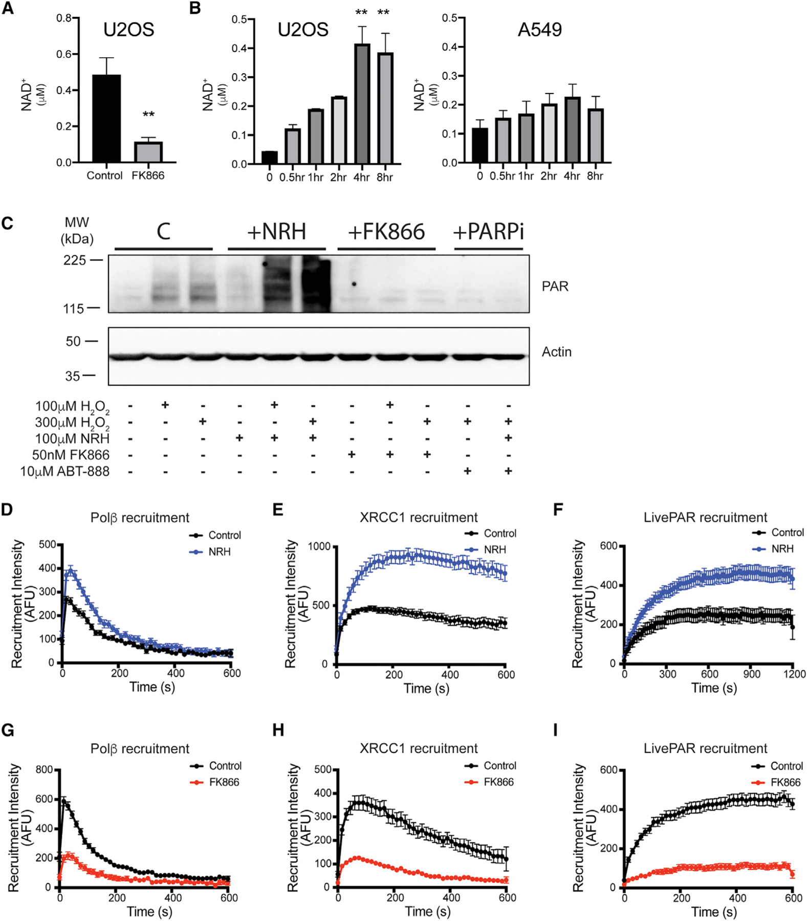
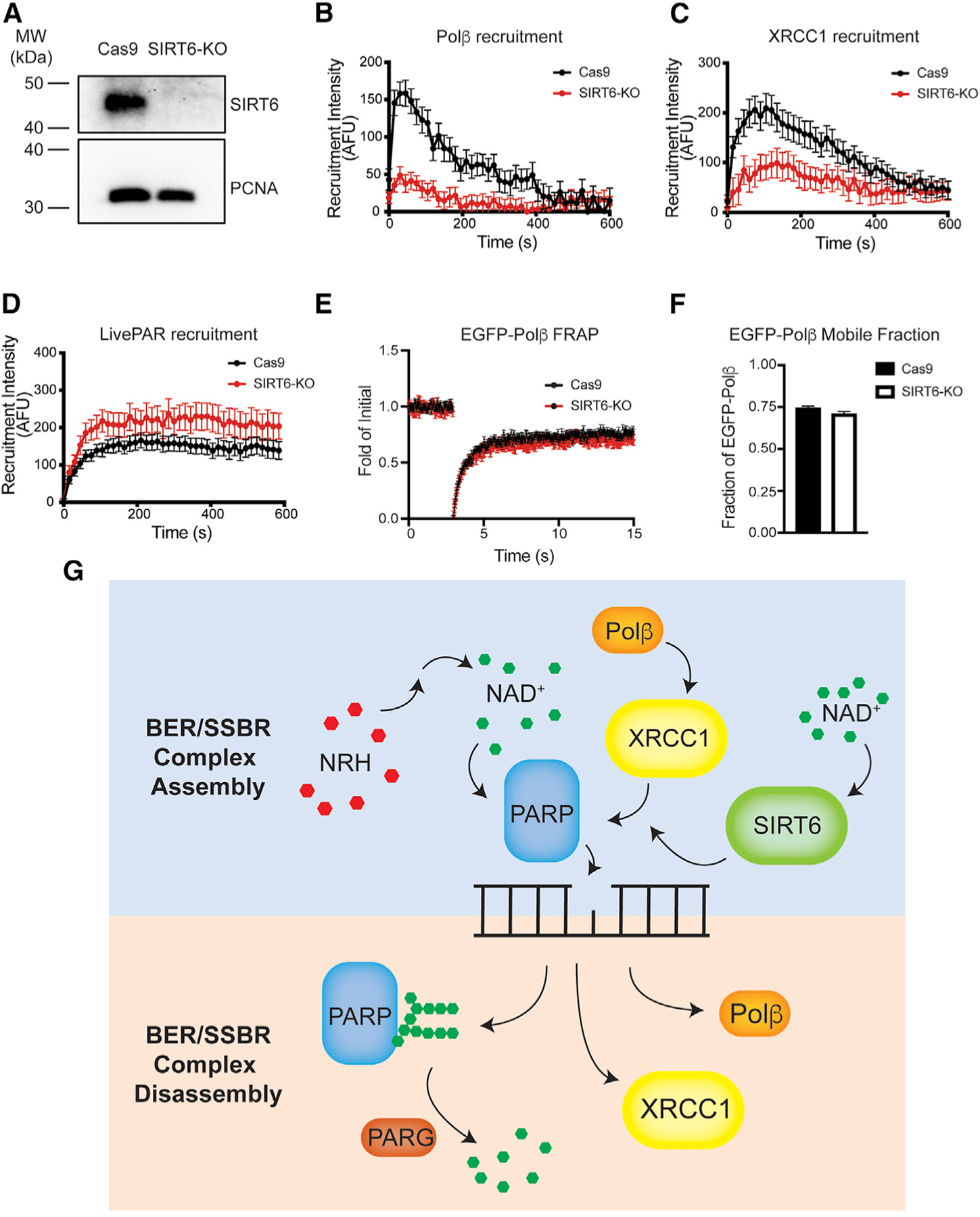
Assembly and disassembly of DNA repair protein complexes at DNA damage sites are essential for maintaining genomic integrity. Investigating factors coordinating assembly of the base excision repair (BER) proteins DNA polymerase β (Polβ) and XRCC1 to DNA lesion sites identifies a role for Polβ in regulating XRCC1 disassembly from DNA repair complexes and, conversely, demonstrates Polβ's dependence on XRCC1 for complex assembly. LivePAR, a genetically encoded probe for live-cell imaging of poly(ADP-ribose) (PAR), reveals that Polβ and XRCC1 require PAR for repair-complex assembly, with PARP1 and PARP2 playing unique roles in complex dynamics. Further, BER complex assembly is modulated by attenuation/augmentation of NAD+ biosynthesis. Finally, SIRT6 does not modulate PARP1 or PARP2 activation but does regulate XRCC1 recruitment, leading to diminished Polβ abundance at sites of DNA damage. These findings highlight coordinated yet independent roles for PARP1, PARP2, and SIRT6 and their regulation by NAD+ bioavailability to facilitate BER.

Keywords: BER; DNA polymerase β; LivePAR; NAD(+); NRH; PAR; SIRT6; SSBR; XRCC1; poly(ADP-ribose).


Conflict of interest statement

Declaration of interests R.W.S. is a scientific consultant for Canal House Biosciences, but this company was not involved in this study, nor was the consulting work related to this study. The authors declare no competing interests.

Figures

Figure 1.
Figure 1.. Laser-induced micro-irradiation: Polβ, XRCC1, and LivePAR
(A) Model for Polβ/XRCC1/PAR complex formation. (B) Recruitment of EGFP-Polβ in U2OS cells. (C) Recruitment of XRCC1-EGFP in U2OS cells. (D) Model of LivePAR’s mode of action. LivePAR contains EGFP fused to a poly(ADP-ribose) (PAR)-binding motif that binds to the iso-ADP-ribose moiety (shown in red). (E) Recruitment of LivePAR in U2OS cells. (F) Recruitment of LivePAR and LivePAR(Y107A) in A549 cells. (G) Inhibition of PARP1/PARP2 or PARG alters LivePAR recruitment to sites of laser micro-irradiation in A549 cells. (H) Time to peak recruitment intensity of Polβ, XRCC1, and LivePAR in U2OS cells. (I) Half-life of recruitment of Polβ, XRCC1, and LivePAR in U2OS cells. N.D., not detected. (J) Serial micro-irradiation of EGFP-Polβ in U2OS cells. (K) Serial micro-irradiation of XRCC1-EGFP in U2OS cells. (L) Serial micro-irradiation of LivePAR in U2OS cells. For (B), (C), and (E)–(L), error bars indicate standard error of the mean, n ≥ 35. All laser micro-irradiation was performed at 355 nm. See Figures S1–S3.
Figure 2.
Figure 2.. Overexpression of EGFP-Polβ recruits similarly to endogenously tagged EGFP-Polβ
(A) Genomic editing strategy to target the POLΒ gene in A549 cells. EGFP cDNA was inserted in-frame with the transcriptional start site of POLΒ, and a silent mutation was placed at the PAM site to prevent re-cleavage by Cas9. (B) Allele sequencing results. Of the three alleles in A549 cells, one was not modified, one was modified with the full-length EGFP in-frame with POLΒ exon 1, and one allele displayed a partial 45-bp insertion. Full sequencing results are in Table S2. (C) Immunoblot of A549 and endogenously tagged A549 cells. (D) Spectrally unmixed image of endogenously tagged EGFP-Polβ in A549 cells. Foci in the image demonstrates EGFP-Polβ recruitment. Scale bar denotes 10 mm distance. (E) Recruitment of endogenous EGFP-Polβ (open circles) and overexpressed EGFP-Polβ (closed circles). (F) Time to peak recruitment intensity of endogenous EGFP-Polβ and overexpressed EGFP-Polβ following micro-irradiation. No significant difference was observed (Student’s t test). (G) Half-life of recruitment of endogenous EGFP-Polβ and overexpressed EGFP-Polβ following micro-irradiation. A significant difference (p < 0.05) was observed (Student’s t test). For (E)–(G), error bars indicate standard error of the mean, n R 35. All laser micro-irradiation was performed at 355 nm. See Figure S4.
Figure 3.
Figure 3.. Loss of Polβ enzymatic activity does not alter its recruitment kinetics
(A) Recruitment of EGFP-Polβ, dRP lyase mutant EGFP-Polβ(K72A), and polymerase mutant EGFP-Polβ(D256A) in U2OS cells. Cells retained endogenous Polβ. (B) Time to peak recruitment intensity of EGFP-Polβ, EGFP-Polβ(K72A), and EGFP-Polβ(D256A) in U2OS cells. (C) Half-life of recruitment of EGFP-Polβ, EGFP-Polβ(K72A), and EGFP-Polβ(D256A). (D) Immunoblots of Polβ, XRCC1, and PCNA of whole-cell protein lysates prepared from U2OS/Cas9 and two separate U2OS/POLΒ-KO cells, generated using two different guide RNAs. (E) Recruitment of EGFP-Polβ, dRP lyase mutant EGFP-Polβ(K72A), and polymerase mutant EGFP-Polβ(D256A) in U2OS/POLΒ-KO(1.7). (F) Recruitment of EGFP-Polβ and dRP lyase triple mutant EGFP-Polβ(K35A/K68AK72A) in U2OS/POLΒ-KO(1.7). For (A)–(C), (E), and (F), error bars indicate standard error of the mean, n ≥ 35. All laser micro-irradiation was performed at 355nm. See Figure S5.
Figure 4.
Figure 4.. Recruitment of Polβ is dependent on XRCC1, while Polβ enables XRCC1 complex dissociation
(A) Immunoblots of XRCC1 and PCNA of whole-cell protein lysates prepared from U2OS/Cas9 and two separate U2OS/XRCC1-KO cells, generated using two different guide RNAs. (B) Recruitment of EGFP-Polβ when expressed in XRCC1-KO cells with and without PARG inhibition (PDD00017273). (C) Recruitment of EGFP-Polβ(WT) and the XRCC1-binding-deficient triple-mutant EGFP-Polβ(L301R/V303R/V306R, TM) when expressed in A549 cells. (D) Recruitment of XRCC1-EGFP when expressed in U2OS/POLΒ-KO cells. (E) Recruitment of XRCC1-EGFP when expressed in U2OS/POLΒ-KO cells with Polβ expression restored. (F) Recruitment of LivePAR when expressed in U2OS/POLΒ-KO cells. (G) Recruitment of EGFP-LIG3 in U2OS/Cas9, U2OS/POLΒ-KO(1.7), and U2OS/XRCC1-KO(E2) cells or following PARP inhibition (ABT-888). For (B)–(G), error bars indicate standard error of the mean, n ≥ 35. All laser micro-irradiation was performed at 355 nm. See Figures S6–S8.
Figure 5.
Figure 5.. Polβ and XRCC1 complex dynamics are dependent on PAR
(A) Immunoblot of PARP1 in U2OS/Cas9 and U2OS/PARP1-KO cells. (B) Recruitment of EGFP-Polβ in U2OS/PARP1-KO cells. (C) Recruitment of EGFP-Polβ in U2OS, U2OS/PARP2-KO or U2OS/PARP1-KO/PARP2-KO cells. (D) Recruitment of EGFP-Polβ in A549 cells following PARP or PARG inhibition. (E) Recruitment of XRCC1-EGFP in A549 cells following PARP or PARG inhibition. For (B)–(E), error bars indicate standard error of the mean, n ≥ 35. All laser micro-irradiation was performed at 355 nm. See Figure S9.
Figure 6.
Figure 6.. Polβ and XRCC1 complex dynamics are regulated by NAD+ bioavailability
(A) NAD+ concentrations in U2OS cells following treatment with FK866, n = 12. FK866 reduced NAD+ concentrations (**p < 0.01; Student’s t test). (B) Time course of NAD+ levels in U2OS cells following NRH treatment (100 µM), n = 6. NRH increased cellular NAD+ concentrations in U2OS cells (**p < 0.01; one-way ANOVA, Tukey post hoc test), but not in A549 cells. (C) Immunoblots of PAR formation in U2OS cells with H2O2 alone (100 µM or 300 µM, 15 min) or following pre-treatments of NRH (100 µM, 4 h), FK866 (50 nM, 24 h), or ABT-888 (10 mM, 1 h). (D) Recruitment of EGFP-Polβ in U2OS cells following NRH treatment. (E) Recruitment of XRCC1-EGFP in U2OS cells following NRH treatment. (F) Recruitment of LivePAR in U2OS cells following NRH treatment. (G) Recruitment of EGFP-Polβ in U2OS cells following FK866 treatment. (H) Recruitment of XRCC1-EGFP in U2OS cells following FK866 treatment. (I) Recruitment of LivePAR in U2OS cells following FK866 treatment. For (A), (C), (G)–(I), FK866 treatment was 50 nM for 24 h. For (D)–(F), NRH treatment was 100 µM for 4 h. For (A), (B), (D)–(I), error bars indicate standard error of the mean. For (D)–(I), n ≥ 35. All laser micro-irradiation was performed at 355 nm. See Figures S10 and S11.
Figure 7.
Figure 7.. Loss of SIRT6 impairs Polβ and XRCC1 complex assembly, but not PAR formation
(A) Immunoblot of SIRT6 in A549/Cas9 and A549/SIRT6-KO cells. (B) Recruitment of EGFP-Polβ in A549/Cas9 and A549/SIRT6-KO cells. (C) Recruitment of XRCC1-EGFP in A549/Cas9 and A549/SIRT6-KO cells. (D) Recruitment of LivePAR in A549/Cas9 and A549/SIRT6-KO cells. (E) Fluorescence recovery after photobleaching (FRAP) traces for EGFP-Polβ in A549/Cas9 or A549/SIRT6-KO cells. (F) FRAP-derived mobile fraction of EGFP-Polβ protein in A549/Cas9 or A549/SIRT6-KO cells. No significant difference was observed (Student’s t test). (G) Model depicting the impact of SIRT6 on PAR-dependent recruitment of XRCC1 to sites of DNA damage. PARPs initiate BER/SSBR complex assembly following micro-irradiation, with PAR formation unchanged in SIRT6-KO cells. SIRT6 regulates recruitment of XRCC1 to PAR following micro-irradiation. Reduced XRCC1 recruitment in SIRT6-KO cells reduces the recruitment of XRCC1 binding proteins such as Polβ. BER/SSBR complex disassembly appears unaffected. For (A)–(F), error bars indicate standard error of the mean, n ≥ 35. All laser micro-irradiation was performed at 355 nm. See Figure S12.

Similar articles

Cited by

References

    1. Abbotts R, and Wilson DM 3rd. (2017). Coordination of DNA single strand break repair. Free Radic. Biol. Med 107, 228–244. - PMC - PubMed
    1. Almeida KH, and Sobol RW (2007). A unified view of base excision repair: lesion-dependent protein complexes regulated by post-translational modification. DNA Repair (Amst.) 6, 695–711. - PMC - PubMed
    1. Beard BC, Wilson SH, and Smerdon MJ (2003). Suppressed catalytic activity of base excision repair enzymes on rotationally positioned uracil in nucleosomes. Proc. Natl. Acad. Sci. USA 100, 7465–7470. - PMC - PubMed
    1. Breslin C, Hornyak P, Ridley A, Rulten SL, Hanzlikova H, Oliver AW, and Caldecott KW (2015). The XRCC1 phosphate-binding pocket binds poly (ADP-ribose) and is required for XRCC1 function. Nucleic Acids Res 43, 6934–6944. - PMC - PubMed
    1. Chiarugi A, Dӧlle C, Felici R, and Ziegler M (2012). The NAD metabolome–a key determinant of cancer cell biology. Nat. Rev. Cancer 12, 741–752. - PubMed

Publication types

MeSH terms