

Anterior skull base reconstruction using an anterolateral thigh free flap
- PMID: 34732034
- PMCID: PMC8568499
- DOI: 10.7181/acfs.2021.00290
Anterior skull base reconstruction using an anterolateral thigh free flap
Abstract
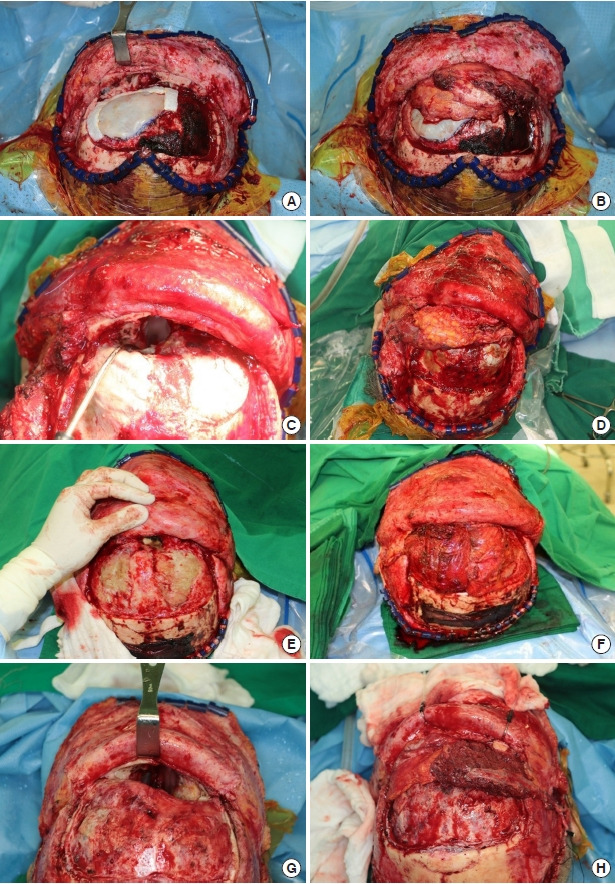
Background: Galeal or temporalis muscle flaps have been traditionally used to reconstruct skull base defects after tumor removal. Unfortunately, these flaps do not provide sufficient vascularized tissue for a dural seal in extensive defects. This study describes the successful coverage of large skull base defects using anterolateral thigh (ALT) free flaps.
Methods: This retrospective study included five patients who underwent skull base surgery between June 2018 and June 2021. Reconstruction was performed using an ALT free flap to cover defects that included the intracranial space and extended to the frontal sinus and cribriform plate.
Results: There were no major complications, such as ascending infections or cerebrospinal leakage. Postoperative magnetic resonance imaging showed that the flaps were well-maintained in all patients.
Conclusion: Successful reconstruction was performed using ALT free flaps for large anterior skull base defects. In conclusion, the ALT free flap is an effective option for preventing communication between the nasal cavity and the intracranial space.
Keywords: Free tissue flaps; Reconstructive surgery; Temporal artery.
Conflict of interest statement
No potential conflict of interest relevant to this article was reported.
Figures


Similar articles
-
Huge Anterior Skull Base Defect Reconstruction on Communicating Between Cranium and Nasal Cavity: Combination Flap of Galeal Flap and Reverse Temporalis Flap.J Craniofac Surg. 2020 Mar/Apr;31(2):436-439. doi: 10.1097/SCS.0000000000006221. J Craniofac Surg. 2020. PMID: 32049922
-
Reconstruction of Complex Lateral Skull Base Defects After Oral Cancer Resection With Individualized Anterolateral Thigh Flap.Front Oncol. 2021 Sep 22;11:743370. doi: 10.3389/fonc.2021.743370. eCollection 2021. Front Oncol. 2021. PMID: 34631580 Free PMC article.
-
Extended temporalis flap for skull base reconstruction.Arch Craniofac Surg. 2019 Apr;20(2):126-129. doi: 10.7181/acfs.2018.02278. Epub 2019 Apr 20. Arch Craniofac Surg. 2019. PMID: 31048650 Free PMC article.
-
Free tissue reconstruction of the anterior skull base: A review.World J Otorhinolaryngol Head Neck Surg. 2020 Apr 17;6(2):132-136. doi: 10.1016/j.wjorl.2020.01.004. eCollection 2020 Jun. World J Otorhinolaryngol Head Neck Surg. 2020. PMID: 32596659 Free PMC article. Review.
-
Reconstruction of Skull Base Defects.Atlas Oral Maxillofac Surg Clin North Am. 2025 Mar;33(1):69-79. doi: 10.1016/j.cxom.2024.08.004. Epub 2024 Oct 2. Atlas Oral Maxillofac Surg Clin North Am. 2025. PMID: 39929560 Review. No abstract available.
Cited by
-
Current concepts of neurofibromatosis type 1: pathophysiology and treatment.Arch Craniofac Surg. 2022 Feb;23(1):6-16. doi: 10.7181/acfs.2022.00633. Epub 2022 Feb 20. Arch Craniofac Surg. 2022. PMID: 35255591 Free PMC article.
-
Adipofascial anterolateral thigh free flap in head and neck reconstruction: a case series.J Laryngol Otol. 2025 Mar;139(3):237-241. doi: 10.1017/S0022215124001385. J Laryngol Otol. 2025. PMID: 39428596 Free PMC article.
-
[Application of iceberg theory in the diagnosis and treatment of refractory wounds at the surgical site after craniotomy].Zhonghua Shao Shang Yu Chuang Mian Xiu Fu Za Zhi. 2025 Jun 20;41(6):509-515. doi: 10.3760/cma.j.cn501225-20250119-00028. Zhonghua Shao Shang Yu Chuang Mian Xiu Fu Za Zhi. 2025. PMID: 40588399 Free PMC article. Chinese.
-
Perforating patterns of cutaneous perforator vessels in anterolateral thigh flaps for head and neck reconstruction and clinical outcomes.Arch Craniofac Surg. 2022 Apr;23(2):64-70. doi: 10.7181/acfs.2022.00171. Epub 2022 Apr 20. Arch Craniofac Surg. 2022. PMID: 35526841 Free PMC article.
References
-
- Gullane PJ, Lipa JE, Novak CB, Neligan PC. Reconstruction of skull base defects. Clin Plast Surg. 2005;32:391–9. - PubMed
-
- Chang DW, Langstein HN, Gupta A, De Monte F, Do KA, Wang X, et al. Reconstructive management of cranial base defects after tumor ablation. Plast Reconstr Surg. 2001;107:1346–57. - PubMed
-
- Menderes A, Yilmaz M, Vayvada H, Demirdover C, Barutcu A. Reverse temporalis muscle flap for the reconstruction of orbital exenteration defects. Ann Plast Surg. 2002;48:521–6. - PubMed
Grants and funding
LinkOut - more resources
Full Text Sources
