. 2022 Jan;33(1):238-252.
doi: 10.1681/ASN.2021060794. Epub 2021 Nov 3.

Discovery of Autoantibodies Targeting Nephrin in Minimal Change Disease Supports a Novel Autoimmune Etiology

Affiliations

Discovery of Autoantibodies Targeting Nephrin in Minimal Change Disease Supports a Novel Autoimmune Etiology

Andrew J B Watts et al. J Am Soc Nephrol. 2022 Jan.

Abstract

Background: Failure of the glomerular filtration barrier, primarily by loss of slit diaphragm architecture, underlies nephrotic syndrome in minimal change disease. The etiology remains unknown. The efficacy of B cell-targeted therapies in some patients, together with the known proteinuric effect of anti-nephrin antibodies in rodent models, prompted us to hypothesize that nephrin autoantibodies may be present in patients with minimal change disease.

Methods: We evaluated sera from patients with minimal change disease, enrolled in the Nephrotic Syndrome Study Network (NEPTUNE) cohort and from our own institutions, for circulating nephrin autoantibodies by indirect ELISA and by immunoprecipitation of full-length nephrin from human glomerular extract or a recombinant purified extracellular domain of human nephrin. We also evaluated renal biopsies from our institutions for podocyte-associated punctate IgG colocalizing with nephrin by immunofluorescence.

Results: In two independent patient cohorts, we identified circulating nephrin autoantibodies during active disease that were significantly reduced or absent during treatment response in a subset of patients with minimal change disease. We correlated the presence of these autoantibodies with podocyte-associated punctate IgG in renal biopsies from our institutions. We also identified a patient with steroid-dependent childhood minimal change disease that progressed to end stage kidney disease; she developed a massive post-transplant recurrence of proteinuria that was associated with high pretransplant circulating nephrin autoantibodies.

Conclusions: Our discovery of nephrin autoantibodies in a subset of adults and children with minimal change disease aligns with published animal studies and provides further support for an autoimmune etiology. We propose a new molecular classification of nephrin autoantibody minimal change disease to serve as a framework for instigation of precision therapeutics for these patients.

Keywords: autoantibodies; glomerular disease; glomerular filtration barrier; idiopathic nephrotic syndrome; immunology; nephrin; pathology; podocyte; proteinuria.


Figures

None
Graphical abstract
Figure 1.
Figure 1.
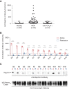
Circulating autoantibodies against nephrin are present in a subset of patients with MCD from the NEPTUNE study cohort and correlate with disease activity. (A) Antibodies against the extracellular domain of recombinant human nephrin (hNephrinG1059) were measured by indirect ELISA. Antigen-specific binding was determined by subtracting the average OD450nm of duplicate uncoated wells (nonspecific background) from the average OD450nm of duplicate hNephrinG1059 coated wells for each individual patient sample. A relative antibody titer was then determined from a standard curve that was generated using a single positive patient sample with a 1:100 dilution defined as containing 1000 U/ml. The threshold for anti-nephrin antibody positivity (187 U/ml) was defined as the maximum antibody titer in a healthy control population (n=30) without kidney disease, as the cohort was not normally distributed and to maximize specificity. The earliest serum sample available during active disease (UPCR >3 g/g on the day of sample collection) was positive for anti-nephrin antibodies in 18 (29%) of 62 patients with biopsy proven MCD from the NEPTUNE cohort. Fifty-three (98%) of 54 nephrotic control patients with anti-hPLA2R antibodies, as determined by clinical ELISA and IIFT assays (Euroimmun), were negative for anti-nephrin antibodies. The intra- and inter-assay coefficient of variances for the anti-nephrin antibody ELISA were 5.56% and 14.36%, respectively. The antibody titers for the NEPTUNE patients and controls are given in Supplemental Table 1. (B) Eleven of the 18 NEPTUNE patients who were anti-nephrin antibody positive during active disease (blue bar) had a subsequent serum sample available during complete remission (UPCR <0.3 g/g on the day of sample collection) which was anti-nephrin antibody negative (red bar) in all patients. Dotted line indicates threshold for positive antibody titer (187 U/ml). The t test was used to compare differences between the active and remission samples, **P<0.01; ***P<0.001. (C) The same serum samples evaluated by ELISA from the NEPTUNE cohort (B) were evaluated for their ability to immunoprecipitate nephrin from HGE derived from non-diseased human kidnies. In keeping with the ELISA results, only serum obtained during active disease (indicated by an arrow with an “A” [blue colored] above it) immunoprecipitated nephrin, whereas serum obtained during remission (indicated by an arrow with an “R” [red colored] above it) did not. Total IgG was comparable between active and remission samples for each patient.
Figure 2.
Figure 2.
Kaplan–Meier estimates for complete remission (CR) and relapse-free period in the NEPTUNE cohort. (A) Time to CR from enrollment was similar between the anti-nephrin antibody (Ab) positive (median time 4.4 months) and negative groups (median time 5.4 months) by a log-rank test (P=0.72). (B) The relapse-free period following CR was shorter in the anti-nephrin antibody positive group (median time 6 months) compared with the antibody negative group (median time 21.57 months), although this finding did not reach statistical significance by a log-rank test (P=0.09). CR was defined as UPCR <0.3 g/g. Relapse when UPCR >3 g/g after reaching CR.
Figure 3.
Figure 3.
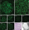
Routine clinical epifluorescence microscopy, periodic-acid Schiff (PAS) stained light microscopy and electron microscopy (EM) images of IgG-positive MCD (MCD7+). Top row: The diffuse punctate staining seen with FITC-conjugated IgG antibody is not seen by albumin staining. Note the positive albumin staining in proximal tubule reabsorption droplets. Middle row: Staining for IgG subtypes confirms no restriction of the punctate staining, and, in this case, more immunoreactivity for IgG1 and IgG2 compared with IgG3 and IgG4 subtypes was observed. Bottom row: Staining for κ and λ light chains shows equal intensity and an appearance mirroring the IgG staining. PAS staining shows minimal light microscopic changes and EM demonstrates diffuse podocyte foot process effacement. Scale bars: Immunofluorescence and PAS: 20 μm. Scale bar: EM: 1 μm.
Figure 4.
Figure 4.
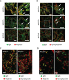
Punctate IgG present in a subset of MCD renal biopsies colocalizes with the critical podocyte slit diaphragm protein nephrin. (A) Representative confocal microscopy images of glomeruli in IgG-positive MCD (MCD1+/MCD7+) and IgG-negative MCD (MCD5−), stained for IgG (green) and the podocyte slit diaphragm protein nephrin (red). There is a clear overlap (yellow) of the punctate IgG (not the background) with nephrin in the IgG-positive MCD+ biopsies (white arrows) but not in IgG-negative MCD biopsies.The right panel shows magified images of the boxed areas for each corresponding image in the left panel. Scale bar: 10 µm. (B) Representative confocal microscopy images of glomeruli in IgG-positive MCD (MCD1+/MCD7+) and IgG-negative MCD (MCD5−), stained for IgG (green) and the podocyte cytoskeletal protein synaptopodin (red). There is no discernable overlap between the punctate IgG (indicated by white arrows) and synaptopodin in any of those patients. Magnified images for each boxed area in the biopsies are shown in the corresponding panel on the right. . Scale bar: 10 µm. (C) Super-resolution SIM images of 0.125µm individual Z-slices showing en face views of the podocyte junction from a representative IgG-positive MCD renal biopsy (MCD1+) in which the nephrin remains glomerular basement membrane–associated, forming a curvilinear pattern. The left image shows colocalization (yellow) of IgG (green) with the slit diaphragm protein nephrin (red), in contrast to mutual exclusivity with the foot process–associated synaptopodin (red) shown in the right image, indicating intimate spatial association with nephrin along the podocyte slit diaphragm. (Full image stack is shown in Supplemental Figure 6.) Scale bar: 1 µm. (D) Super-resolution SIM images of 0.125µm individual Z-slices from a representative IgG-positive MCD renal biopsy (MCD7+) in which nephrin is redistributed to a more granular pattern. The left image shows colocalization (yellow) of IgG (green) with the slit diaphragm protein nephrin (red), in contrast to mutual exclusivity with the foot process–associated synaptopodin (red) shown in the right image, indicating a continued close spatial association of the IgG with the redistributed nephrin. (Full image stack is shown in Supplemental Figure 6.) Scale bar: 1 µm.
Figure 5.
Figure 5.
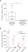
Circulating autoantibodies against nephrin are exclusively present in patients with renal biopsy IgG-positive MCD. (A) All of the patients with MCD and IgG deposition on biopsy (n=9) were serologically anti-nephrin antibody positive, whereas all of the control subjects lacking IgG deposition on biopsy, consisting of diabetic nephropathy (n=2), amyloidosis (n=1), IgG-negative FSGS (n=2), IgG-negative TL (n=1), normal (n=1), disease-free region of tumor nephrectomy (n=2), and IgG-negative MCD (n=3), were serologically anti-nephrin antibody negative (n=12). The Mann–Whitney U test was used to compare differences between the groups, ***P<0.001. (B) Serum/plasma samples were obtained from patients with biopsy proven IgG-positive MCD (MCD+) during active disease (within 7 days of presentation with nephrotic syndrome) and follow-up samples were obtained during complete (MCD4+, MCD7+) or partial (MCD8+) remission on the day of sample collection. For MCD3+, the follow-up serum sample was obtained approximately 3 weeks after entering a period of sustained complete remission. The threshold for a positive anti-nephrin antibody titer of 187 U/ml (indicated by dotted line) was based on the upper limit of a healthy control population. Anti-nephrin antibodies in serum/plasma were undetectable or significantly reduced to below the threshold for positivity (red bar) during clinical remission compared with those during active disease (blue bar). Complete remission was defined as UPCR <0.3 g/g or urinary albumin creatinine ratio <0.2 g/g. Partial remission was defined as a >50% reduction in proteinuria (UPCR) that did not fall below 0.3 g/g. The t test was used to compare differences between the active and remission samples, **P<0.01; ***P<0.001.
Figure 6.
Figure 6.
Pretransplant nephrin autoantibodies are associated with early post-transplant massive proteinuria recurrence in a patient with steroid-dependent MCD that progressed to ESKD. (A) Clinical course of patient with childhood onset, steroid-dependent MCD that progressed to ESKD with subsequent biopsies showing FSGS. She developed early, massive proteinuria recurrence, following a cadaveric, pediatric donor kidney transplant that rapidly responded to treatment with plasmapheresis (purple lines) and rituximab (green lines) shown above the graph. Full clinical details are available in the Supplemental Clinical Case. (B) Two pretransplant serum samples (day −617, day −528) and the initial plasmapheresate (shaded box) on day 13 (d13 (PP), indicated by arrow in (A)) tested positive by ELISA for anti-nephrin antibodies (dotted line indicates threshold for positivity of 187 U/ml). Serum samples evaluated following treatment response at day 27 (d27, indicated by arrow in (A)) and day 365 (UPCR <0.3 g/g) were negative for anti-nephrin antibodies. (C) Similarly, the pretransplant serum and plasmapheresate immunoprecipitated nephrin from HGE (derived from disease-free human kidneys) in keeping with the ELISA findings.

Comment in

References

    1. Vivarelli M, Massella L, Ruggiero B, Emma F: Minimal change disease. Clin J Am Soc Nephrol 12: 332–345, 2017 - PMC - PubMed
    1. Grahammer F, Schell C, Huber TB: The podocyte slit diaphragm--from a thin grey line to a complex signalling hub. Nat Rev Nephrol 9: 587–598, 2013 - PubMed
    1. Kestilä M, Lenkkeri U, Männikkö M, Lamerdin J, McCready P, Putaala H, et al. : Positionally cloned gene for a novel glomerular protein--nephrin--is mutated in congenital nephrotic syndrome. Mol Cell 1: 575–582, 1998 - PubMed
    1. Liu L, Doné SC, Khoshnoodi J, Bertorello A, Wartiovaara J, Berggren PO, et al. : Defective nephrin trafficking caused by missense mutations in the NPHS1 gene: Insight into the mechanisms of congenital nephrotic syndrome. Hum Mol Genet 10: 2637–2644, 2001 - PubMed
    1. Elie V, Fakhoury M, Deschênes G, Jacqz-Aigrain E: Physiopathology of idiopathic nephrotic syndrome: Lessons from glucocorticoids and epigenetic perspectives. Pediatr Nephrol 27: 1249–1256, 2012 - PubMed

Publication types