. 2021 Nov 3;11(1):21547.
doi: 10.1038/s41598-021-00931-1.

An open source extrusion bioprinter based on the E3D motion system and tool changer to enable FRESH and multimaterial bioprinting

Affiliations

An open source extrusion bioprinter based on the E3D motion system and tool changer to enable FRESH and multimaterial bioprinting

Adam Engberg et al. Sci Rep. .

Abstract

Bioprinting is increasingly used to create complex tissue constructs for an array of research applications, and there are also increasing efforts to print tissues for transplantation. Bioprinting may also prove valuable in the context of drug screening for personalized medicine for treatment of diseases such as cancer. However, the rapidly expanding bioprinting research field is currently limited by access to bioprinters. To increase the availability of bioprinting technologies we present here an open source extrusion bioprinter based on the E3D motion system and tool changer to enable high-resolution multimaterial bioprinting. As proof of concept, the bioprinter is used to create collagen constructs using freeform reversible embedding of suspended hydrogels (FRESH) methodology, as well as multimaterial constructs composed of distinct sections of laminin and collagen. Data is presented demonstrating that the bioprinted constructs support growth of cells either seeded onto printed constructs or included in the bioink prior to bioprinting. This open source bioprinter is easily adapted for different bioprinting applications, and additional tools can be incorporated to increase the capabilities of the system.


Conflict of interest statement

The authors declare no competing interests.

Figures

Figure 1
Figure 1
Overview of the open source bioprinter based on the E3D motion system and tool changer. (a) Front view and side view CAD illustrations of the open source bioprinter. A transparent polycarbonate box with an integrated HEPA filter with an air intake fan encloses the tool changer to reduce the risk for contamination during the bioprinting process. The tool changer, which is translated along the x- and y-axes, and can retrieve tools from four different tool positions (four syringe pump extrusion tools are shown). The print platform is translated along the z-axis and is equipped with a calibration camera with a microswitch that is used to correct for changes in extrusion position following a change of syringe pump tool. A touch screen attached to the exterior of the cabinet facilitates control of tool selection and calibration functions. (b) Isometric views of the open source bioprinter. The left panel presents a photograph of the assembled bioprinter and the right panel presents a CAD illustration.
Figure 2
Figure 2
Design of the syringe pump extrusion tool. (a) A photograph of the assembled syringe pump extrusion tool. The syringe plunger is pushed down by a lead screw via a plunger press. The lead screw is linearly translated along the z-axis through a stepper motor, which is attached atop the 3D printed plunger housing. 3D printed exchangeable syringe mounts are attached to the base of the plunger housing, and the entire syringe pump extrusion tool is attached to the tool exchanger system via the E3D tool mount fixture. (b) Isometric and exploded views of CAD illustrations of the syringe pump extrusion tool. (c) Linear regression analysis comparing the vertical displacement (mm) of the syringe plunger as measured by a dial indicator with the intended step sizes 10 mm, 1 mm or 0.1 mm set on the stepper motor. Ten measurements were recorded for each step size (red dotted lines indicate S.D.). (d) A differential interference contrast image detailing a section of a collagen string bioprinted using the FRESH protocol through a needle with a cross-sectional diameter of 50 μm. Scatterplot of width measurements recorded from multiple locations along the length of the printed string (100 ± 12 μm).
Figure 3
Figure 3
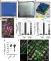
FRESH bioprinting using the open source bioprinter. (a) Design of the construct to be printed using FRESH methodology, and (b) actual FRESH bioprinted construct. (c) 3D rendering of a Col-F stained bioprinted collagen construct based on imaging by confocal microscopy. (d) Example of MDA-MB-231 breast cancer cells seeded onto a bioprinted collagen construct post printing. The cells were stained with SiR-actin and NucBlue to visualize the actin cytoskeleton and cell nuclei respectively. The cells were shown to attach and adopt normal cell morphologies a few hours after cell seeding. The relative level of active apoptosis (e) as measured by Caspase 3/7 activity, and relative occurrence of dead cells (f) as measured by propidium iodide signal in cells grown in glass-bottomed wells or on FRESH bioprinted collagen constructs, in the presence or absence of the apoptosis-inducing toxin staurosporine (10 μM for 5 h) are shown. (g) Analysis of cell viability as assessed by fluorescein diacetate (live) and propidium iodide (dead) staining of MDA-MB-231 breast cancer cells after overnight culture (~ 20 h) on bioprinted collage constructs, and 1 week later. (h) Example of fluorescein diacetate (live cells) and propidium iodide (dead cells) staining of MDA-MB-231 breast cancer cells distributed on the bioprinted collagen constructs following 1 week of culture. Differential interference contrast imaging (DIC) reveals the crisscross patterning of the construct.
Figure 4
Figure 4
Multimaterial bioprinting using the open source bioprinter. (a) Example of fluorescein diacetate (live cells; green) and propidium iodide (dead cells; red) staining of MDA-MB-231 breast cancer cells extruded in laminin bioink directly after extrusion through an 18G needle and following 1 week of culture. (b) Comparative analysis of cell viability following overnight culture (O/N) and one week after extrusion of cells in laminin bioink through an 18 G needle or manually deposited directly from a syringe. (c) Design of the multimaterial construct to be printed using both collagen (green) and cell-laden laminin (red) bioinks, as viewed from above illustrating the print path. (d) Image of the bioprinted multimaterial construct. (e) Col-F staining of the collagen bioink (green) and Celltracker red staining of MDA-MB-231 cells distributed in the laminin bioink.

Similar articles

Cited by

References

    1. Griffith LG, Naughton G. Tissue engineering—Current challenges and expanding opportunities. Science. 2002;295:1009–1014. doi: 10.1126/science.1069210. - DOI - PubMed
    1. Langer R, Vacanti J. Tissue engineering. Science. 1993;260:920–926. doi: 10.1126/science.8493529. - DOI - PubMed
    1. Peng W, et al. 3D bioprinting for drug discovery and development in pharmaceutics. Acta Biomater. 2017;57:26–46. doi: 10.1016/j.actbio.2017.05.025. - DOI - PubMed
    1. Satpathy A, et al. Developments with 3D bioprinting for novel drug discovery. Expert Opin. Drug Discov. 2018;13:1115–1129. doi: 10.1080/17460441.2018.1542427. - DOI - PMC - PubMed
    1. Humphreys BD. Bioprinting better kidney organoids. Nat. Mater. 2021;20:128–130. doi: 10.1038/s41563-020-00881-5. - DOI - PubMed

Publication types