Review
. 2022 Apr;20(4):236-248.
doi: 10.1038/s41579-021-00638-0. Epub 2021 Nov 3.

Physical properties of the bacterial outer membrane

Affiliations
Review

Physical properties of the bacterial outer membrane

Jiawei Sun et al. Nat Rev Microbiol. 2022 Apr.

Abstract

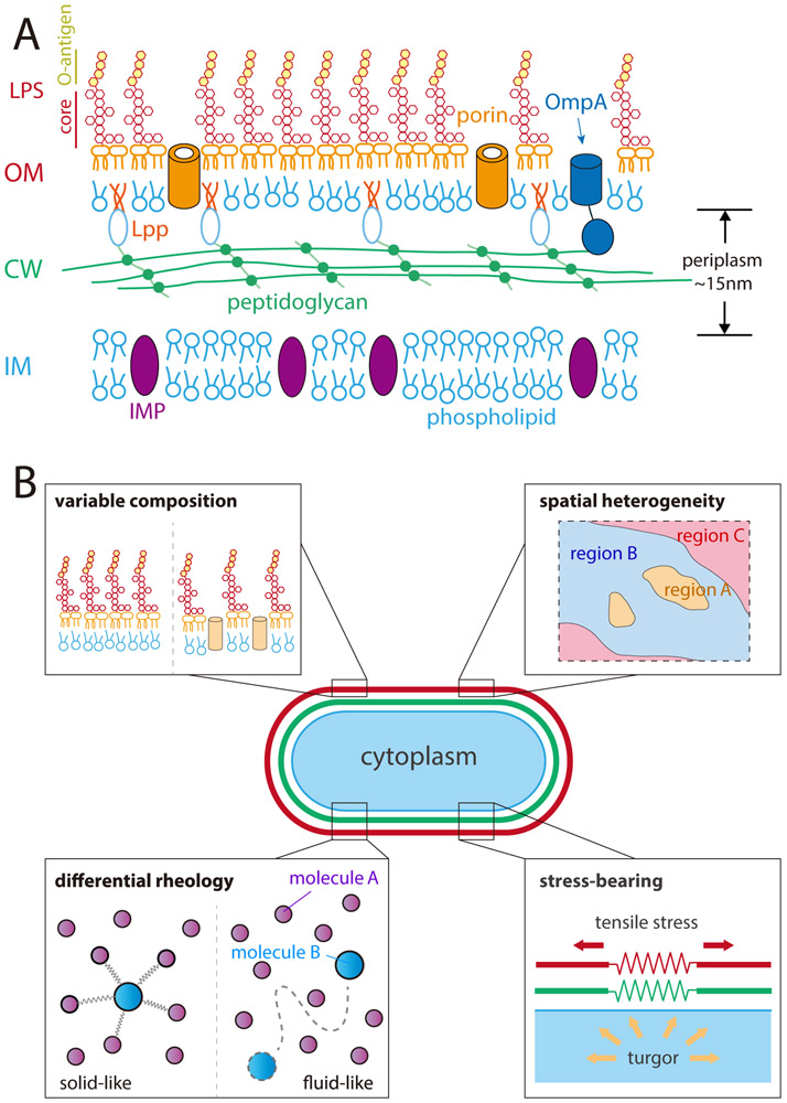
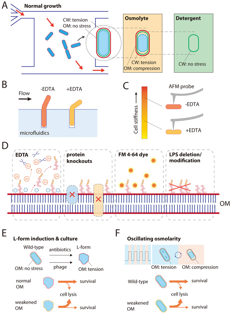
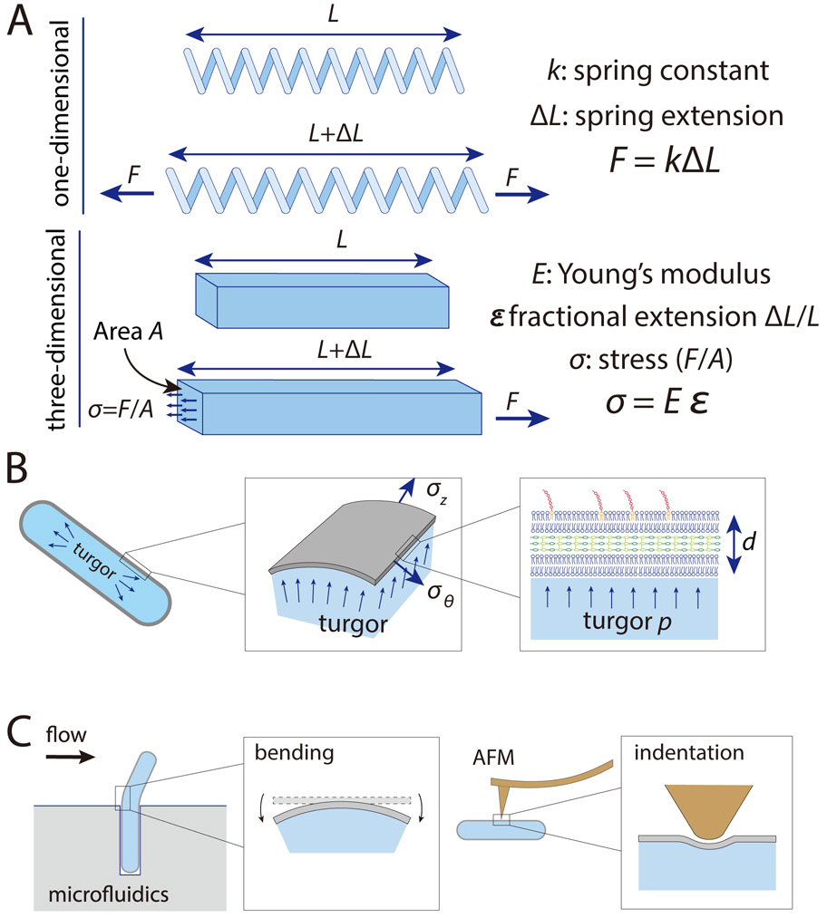
It has long been appreciated that the Gram-negative outer membrane acts as a permeability barrier, but recent studies have uncovered a more expansive and versatile role for the outer membrane in cellular physiology and viability. Owing to recent developments in microfluidics and microscopy, the structural, rheological and mechanical properties of the outer membrane are becoming apparent across multiple scales. In this Review, we discuss experimental and computational studies that have revealed key molecular factors and interactions that give rise to the spatial organization, limited diffusivity and stress-bearing capacity of the outer membrane. These physical properties suggest broad connections between cellular structure and physiology, and we explore future prospects for further elucidation of the implications of outer membrane construction for cellular fitness and survival.


Figures

Figure 1:
Figure 1:. The Gram-negative outer membrane has a diverse makeup and a wide variety of physical and mechanical properties.
A) The cytoplasm of all bacterial cells is surrounded by an inner membrane (IM) composed of phospholipids and inner membrane proteins (IMPs), which is enclosed by the cell wall (CW), a macromolecular network of peptidoglycan (PG) that single-layered in E. coli. In Gram-negative bacteria, the cell wall resides in the periplasm, a ~15-nm thick aqueous compartment enclosed by the inner and outer membranes. The outer membrane (OM) is asymmetric, composed of phospholipids in the inner leaflet and lipopolysaccharides (LPS), along with outer membrane proteins (OMPs) such as porins and lipoproteins such as Lpp, which links the OM and PG. B) The OM (red) can exhibit variable composition, spatial heterogeneity, solid- or fluid-like rheology (diffusive dynamics), and the ability to bear mechanical stresses of similar magnitude to the cell wall (green). As a result, the chemical, physical, and mechanical properties of the OM can have broad impacts on bacterial physiology.
Figure 2:
Figure 2:. Outer membrane rheology and spatial organization are highly distinct from that of the PG or IM.
A) While the PG behaves as a relatively immobile solid due to its highly crosslinked nature, and the IM behaves as a liquid with rapid diffusion of lipids, the OM exhibits limited diffusivity. LPS molecules (purple) and OMPs (red) appear largely immobile, although there is some evidence of diffusive motion in the OM. Green circles represent proteins with relatively high diffusivity. B) At the nanoscale, LPS molecules and OMPs exist in small clusters. At the cellular scale, the density of fluorescently labeled OMPs along the cylindrical portion of the cell is diluted by growth, while the density at the poles remains high. C) At the mesoscale, islands (represented by irregular shapes) including proteins such as the BAM complex expand and spread apart due to insertion of new materials along the cylindrical portion of the cell. After division, clusters at the old pole remain in one daughter cell, while clusters at the new pole have been trapped there by cytokinesis.
Figure 3:
Figure 3:. Probing the magnitude and impact of OM stiffness.
A) In a microfluidic flow cell, exponentially growing E. coli cells are under mechanical stress due to turgor pressure, which is born predominantly by the PG rather than the OM. Cells shrink suddenly when exposed to a hyperosmotic shock due to water efflux, decreasing the overall stress on the cell envelope. In this shrunken state, the cell wall remains partially extended as the OM experiences compression. Removal of the OM by detergent or EDTA treatment allows the cell wall to fully relax to its rest length. B) Growth of E. coli cells initially embedded within a narrow channel leads to exposure of part of the cell body to fluid flow, whose hydrodynamic force can be tuned. EDTA-treated cells deflect more than untreated cells for a given flow strength, highlighting the strength of the OM. C) AFM measurements directly confirm the loss of cell stiffness by EDTA treatment. EDTA-treated cells indented more than untreated cells for the same amount of applied force; several outer membrane mutants also exhibited more indentation. D) In addition to EDTA treatment, OM stiffness can be compromised by deletion of various OM proteins, intercalation of the OM-specific dye FM4-64, or LPS modification such as deletion of the O-antigen. E) E. coli cells adopt a wall-less L-form or wall-deficient spheroplast state when exposed to phage or cell wall-targeting antibiotics, in which the OM bears stress to avoid envelope rupture. F) Cells with compromised OM stiffness are more susceptible to death during osmotic-shock oscillations.
Box Figure:
Box Figure:. The mechanics of cylindrical thin shells.
A) For a one-dimensional spring with spring constant k, the force required to extend the spring by an amount ΔL is F = kΔL. For a three-dimensional material, Young’s modulus E is the analog of the spring constant, and is the ratio of the stress σ to the fractional extension ε. B) The cell envelope of a rod-shaped bacterial cell with cross-sectional radius r can be modeled as a thin shell of thickness d (which is much smaller than r) under turgor pressure p. Along the cylindrical portion the stresses in the axial and circumferential directions are approximately σz=pr2d and σθ=prd. C) Bending in a microfluidic chamber (Figure 3B) and AFM indentation (Figure 3C) explore other modes of deformation such as bending and indentation, respectively.

References

    1. Silhavy TJ, Kahne D & Walker S The bacterial cell envelope. Cold Spring Harb Perspect Biol 2, a000414, doi:10.1101/cshperspect.a000414 (2010). - DOI - PMC - PubMed
    1. Holtje JV Growth of the stress-bearing and shape-maintaining murein sacculus of Escherichia coli. Microbiol Mol Biol Rev 62, 181–203 (1998). - PMC - PubMed
    1. Funahara Y & Nikaido H Asymmetric localization of lipopolysaccharides on the outer membrane of Salmonella typhimurium. Journal of bacteriology 141, 1463–1465 (1980). - PMC - PubMed
    1. Konovalova A & Silhavy TJ Outer membrane lipoprotein biogenesis: Lol is not the end. Philos Trans R Soc Lond B Biol Sci 370, doi:10.1098/rstb.2015.0030 (2015). - DOI - PMC - PubMed
    1. Nikaido H Molecular basis of bacterial outer membrane permeability revisited. Microbiol Mol Biol Rev 67, 593–656 (2003). - PMC - PubMed

Publication types

Substances