Role of 3D Printing and Modeling to Aid in Neuroradiology Education for Medical Trainees
- PMID: 34733071
- PMCID: PMC8560046
- DOI: 10.12788/fp.0134
Role of 3D Printing and Modeling to Aid in Neuroradiology Education for Medical Trainees
Abstract
Background: Applications of 3-dimensional (3D) printing in medical imaging and health care are expanding. Currently, primary uses involve presurgical planning and patient and medical trainee education. Neuroradiology is a complex subdiscipline of radiology that requires further training beyond radiology residency. This review seeks to explore the clinical value of 3D printing and modeling specifically in enhancing neuroradiology education for radiology physician residents and medical trainees.
Methods: A brief review summarizing the key steps from radiologic image to 3D printed model is provided, including storage of computed tomography and magnetic resonance imaging data as digital imaging and communications in medicine files; conversion to standard tessellation language (STL) format; manipulation of STL files in interactive medical image control system software (Materialise) to create 3D models; and 3D printing using various resins via a Formlabs 2 printer.
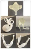
Results: For the purposes of demonstration and proof of concept, neuroanatomy models deemed crucial in early radiology education were created via open-source hardware designs under free or open licenses. 3D-printed objects included a sphenoid bone, cerebellum, skull base, middle ear labyrinth and ossicles, mandible, circle of Willis, carotid aneurysm, and lumbar spine using a combination of clear, white, and elastic resins.
Conclusions: Based on this single-institution experience, 3D-printed complex neuroanatomical structures seem feasible and may enhance resident education and patient safety. These same steps and principles may be applied to other subspecialties of radiology. Artificial intelligence also has the potential to advance the 3D process.
Copyright © 2021 Frontline Medical Communications Inc., Parsippany, NJ, USA.
Conflict of interest statement
Author disclosures The authors report no actual or potential conflicts of interest with regard to this article.
Figures


References
-
- Whyms BJ, Vorperian HK, Gentry LR, Schimek EM, Bersu ET, Chung MK. The effect of computed tomographic scanner parameters and 3-dimensional volume rendering techniques on the accuracy of linear, angular, and volumetric measurements of the mandible. Oral Surg Oral Med, Oral Pathol Oral Radiol. 2013;115(5):682–691. doi: 10.1016/j.oooo.2013.02.008. - DOI - PMC - PubMed
-
- Materialise Cloud. Triangle reduction. [Accessed May 20, 2021]. https://cloud.materialise.com/tools/triangle-reduction.
LinkOut - more resources
Full Text Sources
