. 2021 Oct 18:12:738125.
doi: 10.3389/fmicb.2021.738125. eCollection 2021.

Analysis of the Replication Mechanisms of the Human Papillomavirus Genomes

Affiliations

Analysis of the Replication Mechanisms of the Human Papillomavirus Genomes

Lisett Liblekas et al. Front Microbiol. .

Abstract

The life-cycle of human papillomaviruses (HPVs) includes three distinct phases of the viral genome replication. First, the viral genome is amplified in the infected cells, and this amplification is often accompanied by the oligomerization of the viral genomes. Second stage includes the replication of viral genomes in concert with the host cell genome. The viral genome is further amplified during the third stage of the viral-life cycle, which takes place only in the differentiated keratinocytes. We have previously shown that the HPV18 genomes utilize at least two distinct replication mechanisms during the initial amplification. One of these mechanisms is a well-described bidirectional replication via theta type of replication intermediates. The nature of another replication mechanism utilized by HPV18 involves most likely recombination-dependent replication. In this paper, we show that the usage of different replication mechanisms is a property shared also by other HPV types, namely HPV11 and HPV5. We further show that the emergence of the recombination dependent replication coincides with the oligomerization of the viral genomes and is dependent on the replicative DNA polymerases. We also show that the oligomeric genomes of HPV18 replicate almost exclusively using recombination dependent mechanism, whereas monomeric HPV31 genomes replicate bi-directionally during the maintenance phase of the viral life-cycle.

Keywords: 2D; HPV; non-theta replication; theta replication; virus replication.


Conflict of interest statement

The authors declare that the research was conducted in the absence of any commercial or financial relationships that could be construed as a potential conflict of interest.

Figures
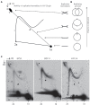
Figure 1
Figure 1
Usage of the non-theta replication mechanism during transient replication is characteristic to different human papillomavirus (HPV) types. (A) Schematic representation of the expected mobility of the theta replication intermediates in neutral-neutral two-dimensional (2D) gel in case a circular replicon is linearized near the origin of replication (ori). Almost a straight diagonal line emanating from the spot of 1n linear molecules represents a doubleY structure, with two replication forks that started from the ori to opposite directions advancing as the line retreats from the spot of 1n linear molecules. A non-linear line ending in the 2D spot represents the structure arising from the non-theta type of replication. (B) Sketch of the HPV replicon at the different stages of bidirectional replication. Ori represents origin of replication; scissors refer to the position, where the HPV genome is linearized. (C) Neutral-neutral 2D analysis of the replication intermediates arising from the HPV5, HPV11, and HPV18 genome replication after transient transfection of the respective genomes into U2OS cells. Episomal DNA was extracted 72h post-transfection, linearized with a restriction enzyme cutting near ori [HPV5 – SacI (nt 702); HPV11- BstEII (nt 7,899) HPV18 – BglI (nt 7,656)] run on an agarose gel at two dimensions and analyzed by Southern blotting (SB). Black arrows refer to the theta replication intermediates, black arrowheads refer to the almost fully replicated genomes, white arrows refer to the non-theta replication intermediates, asterisks depict the linear HPV genome fragments running between the size of 1n and 2n.
Figure 2
Figure 2
Appearance of the non-theta replication intermediates during the transient replication of the HPV18 genomes in the U2OS cells concurs with the oligomerization of the replicated viral genomes. (A) An example of different forms of the HPV18 genome arising as a result of the transient replication in U2OS cells at 3, 4, or 5days after the transfection. Genomic DNA was extracted from the cells at indicated time points, digested with HPV18 non-cutter HindIII and DpnI to separate non-replicated input DNA, resolved on 0.6% gel and analyzed by SB. Migration of the monomeric closed circular (1xCC), monomeric open circular (1XOC), dimeric closed circular (2xCC), and oligomeric forms are shown. Asterisk represents migration of non-replicated, DpnI sensitive fragment of HPV18. (B) Quantification of the replicated monomeric and oligomeric HPV18 genomes at 3, 4, and 5days after the transfection in the U2OS cells. The data are expressed as average means±the SD (*p<0.05; **p<0.01; and ***p<0.001) of four independent experiments. Monomeric open circular and dimeric closed circular genomes migrate together under the conditions used to resolve DNA and were therefore omitted from calculation. (C) An example of different types of the replication intermediates arising during the transient replication of the HPV18 genome in U2OS cells at 3, 4, and 5days post-transfection. Black arrows refer to the theta replication intermediates, black arrowheads refer to the almost fully replicated genomes, white arrows refer to the non-theta replication intermediates. (D) Quantification of the theta- and non-theta replication intermediates arising during the transient replication of the HPV18 genomes at 3, 4, and 5days after the transfection in the U2OS cells. The data are expressed as average means±the SD (*p<0.05; **p<0.01; and ***p<0.001) of four independent experiments.
Figure 3
Figure 3
The non-theta replication of the HPV18 genomes is dependent on the replicative DNA polymerase(s). (A) The cell cycle profile of the aphidicolin arrested cells (0h), followed by the release into the cell cycle at various time points (3, 7.5, and 24h) post-arrest. The U2OS cells were transfected with the HPV18 genome, 48h after the transfection the cells were arrested at G1/S phase using aphidicolin treatment (2μg/ml) for 24h, followed by the removal of the drug and release of the cells to cycling conditions for the indicated periods of time. (B) Neutral-neutral 2D analysis of the replication intermediates arising from the HPV18 genome replication after the transfection into the U2OS cells at various time points after the G1/S block using aphidicolin. Nearly all replication intermediates are absent at the end of 24-h block by aphidicolin (0h). Instead, the collapsed intermediates migrate above the 1n linear molecule spot (indicated by asterisk). At 3h post-release, when majority of the cells have entered the S-phase, both theta and non-theta type of replication intermediates are present. About 7.5h after the release majority of the cells have entered G2/M phase, and still both type of replication intermediates are detectable. At 24h post-release, the cells have restored the normal distribution of the cell cycle and both theta- and non-theta replication intermediates can be seen. Black arrows refer to the theta replication intermediates, black arrowheads refer to the almost fully replicated genomes, and white arrows refer to the non-theta replication intermediates.
Figure 4
Figure 4
Mechanism of replication initiated from the HPV18 ori does not depend on the E1 and E2 protein levels. (A) Neutral-neutral 2D analysis of the replication intermediates arising from the HPV18 E8-genome replication in the presence of growing amounts of the E1 expression vector. (B) Neutral-neutral 2D analysis of the replication intermediates arising from the HPV18 E8-genome replication in the presence of growing amounts of the E2 expression vector. Episomal DNA was extracted 72h after the transfection, linearized with a restriction enzyme BglII cutting near the ori, run on an agarose gel at two dimensions and analyzed by SB. Black arrows refer to the theta replication intermediates, black arrowheads refer to the almost fully replicated genomes, white arrows refer to the non-theta replication intermediates. (C) Western blot analysis showing the expression levels of the HPV18 E1 and E2 proteins, when co-transfected together with the HPV18 E8-genome.
Figure 5
Figure 5
Mechanism of replication initiated from the HPV18 ori depends on the size of the replicon. (A) Transient replication of the HPV18 ori harboring replicons with different sizes in the U2OS cells. pUCURR18 (4kb), pGLURR18 (6kb), or pGEXURR18 (8,2 kb) were transfected together with the HPV18 E1 and E2 expression vectors into the U2OS cells. Episomal DNA was extracted 72h after the transfection, digested with replicon non-cutter (left panel) or single cutter enzyme (right panel), combined with DpnI to digest the non-replicated plasmids, resolved in 0.8% agarose gel and analyzed by SB. Asterisks represent migration of the monomeric closed circular forms of the respective replicons. (B) Neutral-neutral 2D analysis of the replication intermediates arising from the HPV18 harboring replicons of different sizes during the transient replication in the U2OS cells and the SV40 origin containing pCDNA3 replicon in Cos1 cells. The replication intermediate structure that varies between different HPV replicons is indicated with an arrow.
Figure 6
Figure 6
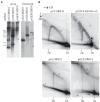
Maintenance replication of the HPV31 genomes proceeds via distinct mechanisms in the CIN612 keratinocytes. (A) SB showing the status of the HPV31 genomes in CIN612 cells. HPV31 noncutter (BamHI) or single cutter (BstXI) enzymes were used to digest episomal DNA. Migration of the monomeric closed circular (1xCC), monomeric open circular (1XOC), linear (1xLin) and oligomeric forms are shown. (B) Neutral-neutral 2D analysis of the replication intermediates arising from the HPV31 maintenance replication in the CIN216 cells, HPV5 and HPV18 maintenance replication in the U2OS cells. Black arrows refer to the theta replication intermediates; white arrows refer to the non-theta replication intermediates.

References

    1. Astell C. R., Chow M. B., Ward D. C. (1985). Sequence analysis of the termini of virion and replicative forms of minute virus of mice DNA suggests a modified rolling hairpin model for autonomous parvovirus DNA replication. J. Virol. 54, 171–177. doi: 10.1128/jvi.54.1.171-177.1985, PMID: - DOI - PMC - PubMed
    1. Auborn K. J., Little R. D., Platt T. H. K., Vaccariello M. A., Schildkraut C. L. (1994). Replicative intermediates of human papillomavirus type 11 in laryngeal papillomas: site of replication initiation and direction of replication. Proc. Natl. Acad. Sci. U. S. A. 91, 7340–7344. doi: 10.1073/pnas.91.15.7340, PMID: - DOI - PMC - PubMed
    1. Bergvall M., Melendy T., Archambault J. (2013). The E1 proteins. Virology 445, 35–56. doi: 10.1016/j.virol.2013.07.020, PMID: - DOI - PMC - PubMed
    1. Borenstein R., Frenkel N. (2009). Cloning human herpes virus 6A genome into bacterial artificial chromosomes and study of DNA replication intermediates. Proc. Natl. Acad. Sci. U. S. A. 106, 19138–19143. doi: 10.1073/pnas.0908504106, PMID: - DOI - PMC - PubMed
    1. Brewer B. J., Fangman W. L. (1987). The localization of replication origins on ARS plasmids in S. cerevisiae. Cell 51, 463–471. doi: 10.1016/0092-8674(87)90642-8, PMID: - DOI - PubMed

LinkOut - more resources