. 2021 Oct 18:12:725325.
doi: 10.3389/fimmu.2021.725325. eCollection 2021.

Interferon Genes Are Influenced by 17β-Estradiol in SLE

Affiliations

Interferon Genes Are Influenced by 17β-Estradiol in SLE

Ram P Singh et al. Front Immunol. .

Abstract

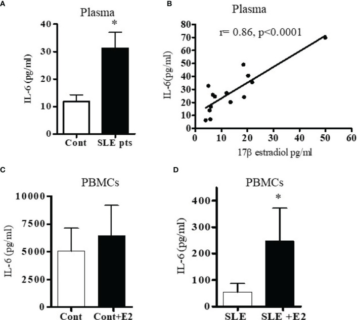
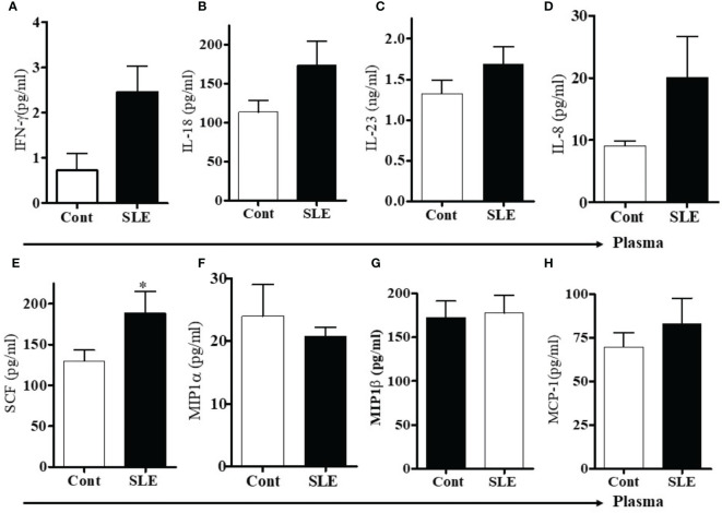
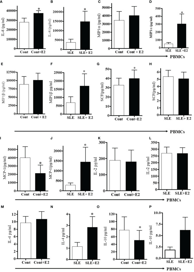
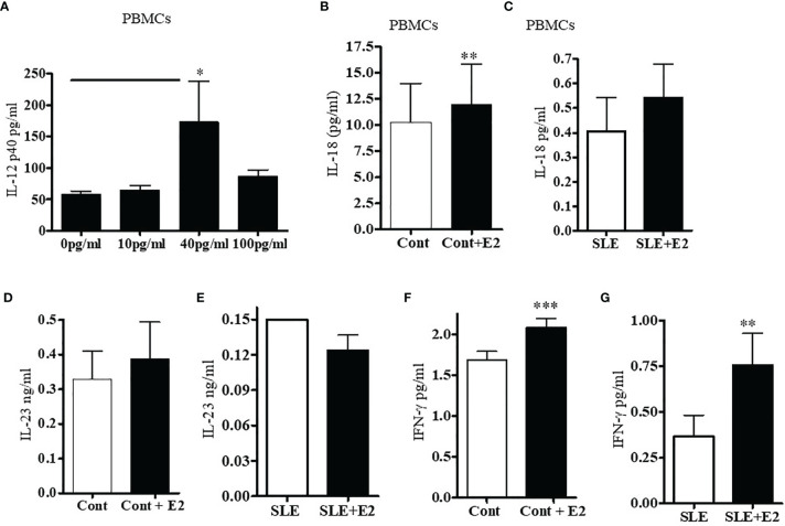
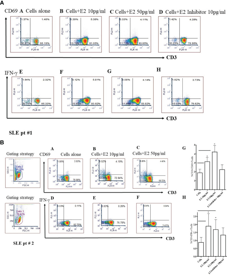
Recent evidence suggests the existence of a nexus between inflammatory pathways and the female sex hormone 17β-estradiol, resulting in increased interferon-stimulated genes (ISGs), autoantibodies, and dysregulation of immune cells in SLE. However, the molecular mechanisms and the effect of estradiol on candidate target genes and their pathways remains poorly understood. Our previous work suggests that female SLE patients have increased estradiol levels compared to healthy controls. In the present study, we explored the effects of 17β-estradiol treatment on expression of IFN (interferons)-stimulated genes and pro-inflammatory cytokines/chemokines. We found significantly increased (5-10-fold) expression of IFN-regulated genes in healthy females. Furthermore, we found significantly increased plasma levels of IL-6, IL-12, IL-17, IL-18, stem cell factor (SCF), and IL-21/IL-23 in SLE patients compared to healthy controls, and those levels positively correlated with the plasma levels of 17β-estradiol. In addition, levels of IL-21 positively correlated with the SLE disease activity index (SLEDAI) score of SLE patients. In vitro treatment of PBMCs from either SLE patients or healthy controls with 17β-estradiol at physiological concentration (~50 pg/ml) also significantly increased secretion of many pro-inflammatory cytokines and chemokines (IL-6, IL-12, IL-17, IL-8, IFN-γ; MIP1α, and MIP1β) in both groups. Further our data revealed that 17β-estradiol significantly increased the percentage of CD3+CD69+ and CD3+IFNγ+ T cells; whereas, simultaneous addition of 17β-estradiol and an ERα inhibitor prevented this effect. Collectively, our findings indicate that 17β-estradiol participates in the induction of pro-inflammatory cytokines and chemokines and further influences interferon genes and pathways.

Keywords: T cells; cytokines/chemokines; estradiol; interferon genes; systemic lupus erythematosus.


Conflict of interest statement

The authors declare that the research was conducted in the absence of any commercial or financial relationships that could be construed as a potential conflict of interest.

Figures

Figure 1
Figure 1
Healthy females have significantly higher interferon-related genes. Peripheral blood mononuclear cells (PBMC) (1-2 x10⁶ cells) were isolated from healthy males (n=5) and females (n=5) and RNA was isolated. 100 ng of RNA was used for real-time PCR analysis of OASL (A) and Ly6E (B). (C) Female BWF1 mice have increased IFI202b gene expression. Splenocytes were obtained from 8-10-week-old male and female BWF1 mice (n=2), RNA isolated, and one-step real-time PCR performed with 100 ng of RNA from each sample. All values were normalized to GAPDH levels. *p < 0.05.
Figure 2
Figure 2
SLE patients have increased levels of the pro-inflammatory cytokine IL-6. Plasma estradiol positively correlates with IL-6. 17β-estradiol increased IL-6 levels in both healthy control and in SLE patients’ PBMCs. (A) Plasma levels of IL-6 were measured in female SLE patients (n=13) and in healthy controls (n=11) by ELISA. (B) Correlation between plasma 17β-estradiol levels and IL-6 in SLE patients (n=14), (p < 0.0001). PBMCs of healthy controls, n=11 (C) and SLE patients, n=11 (D) were cultured with 17β-estradiol (50 pg/ml) for 24-48 h and supernatants measured by multiplex assays. *p < 0.05.
Figure 3
Figure 3
(A) SLE patients have increased levels of IL-17 and IL-21 pro-inflammatory cytokines. Plasma estradiol levels positively correlates with IL-21. (A) IL-21 levels in healthy controls (n=12) vs. SLE patients’ (n=12) plasma. (B) Correlation between plasma estradiol levels and IL-21 in SLE female patients (n=15). (C) Correlation between IL-21 levels and SLEDAI (SLE disease activity index) score in SLE female patients (n=14). (D) IL-17 protein levels in healthy controls (n=12) vs female SLE patients’ (n=12) plasma. (E) IL-17 protein levels measured from supernatant of PBMCs of healthy (n=11) controls PBMCs culture vs healthy controls PBMCs+E2 (50 pg/ml). (F) IL-17 protein levels of SLE (n=11) female patients PBMCs vs SLE patients PBMC+E2 were cultured with 17β-estradiol (50 pg/ml) for 24-48 h and supernatant was measured by multiplex assay (MAP) for IL-17. (G) Correlation between plasma estradiol levels and IL-17 in female SLE patients (n=18). (H) Correlation between plasma estradiol levels and IL-12-p40 in female SLE patients (n=14). *p < 0.05, ***p < 0.001.
Figure 4
Figure 4
Plasma levels of pro-inflammatory cytokines, chemokines including stem cell factor (SCF) were increased in SLE patients. To address the differences of pro-inflammatory cytokines and chemokines between healthy controls and SLE patients, plasma levels of pro-inflammatory cytokines, chemokines and stem cell factor (SCF) were measured in healthy controls (n=10-12) and SLE patients (n= 10-12) by RBM multiplex assay. (A) IFN-γ, (B) IL-18, (C) IL-23, (D) IL-8, (E) Stem cell factor (SCF), (F) MIP1α, (G) MIP1β, and (H) MCP-1. *p < 0.05.
Figure 5
Figure 5
17β-estradiol increases pro-inflammatory chemokines in both healthy control and SLE patients’ PBMCs. PBMCs of female healthy controls and female SLE patients were cultured with 17β-estradiol (50 pg/ml) for 24-48h range (n=10-12). Culture supernatants were obtained and levels of IL-8 (A, B), MIP1α (C, D), MIP1β (E, F), SCF (G, H) MCP-1 (I, J), IL-2 (K, L), IL-4 (M, N), and IL-10 (O, P) were measured by multiplex assays (MAP). Conditions: (A, C, E, G, I, K, M, O). Healthy female control cells (PBMCs), Healthy female control cells + E2 (50 pg/ml) n=11, (B, D, F, H, J, L N, P). SLE female patient cells (PBMCs), SLE female patient cells + E2 (50 pg/ml) n=10. *p < 0.05.
Figure 6
Figure 6
17β-estradiol increases pro-inflammatory cytokines IL-12p40, IL-18, and IL-23 in SLE patients compared to healthy controls. PBMCs from female SLE patients (n=7) were obtained and 2-4x10⁶ cells were cultured with 17β-estradiol (E2 at 10, 40 and 100 pg/ml) for 24-48 h. Culture supernatants were obtained. (A) IL-12p40 levels were measured with ELISA (BioLegend). *p < 0.05. (B) IL-18 levels were measured in the supernatant of healthy control PBMCs treated with E2 (50 pg/ml) (n=11). (C) IL-18 levels were measured in the supernatant of SLE patients PBMCs treated with E2 (50 pg/ml) (n=11). (D) IL-23 levels were measured in the supernatant of healthy control PBMCs treated with E2 (50 pg/ml) (n=8). (E) IL-23 levels were measured in the supernatant of SLE patients PBMCs treated with E2 (50 pg/ml) (n=9). (F) PBMCs (4x10⁶) of healthy controls (n=9) were cultured with 17β-estradiol (50 pg/ml). Culture supernatants were obtained after (24-48 h) and the level of secreted IFNγ was measured with multiplex assay (RBM-MAP, Austin, TX, USA). *p < 0.05. (G) IFNγ levels were measured in the supernatant of SLE patients PBMCs treated with E2 (50 pg/ml) (n=8). *p < 0.05, **p < 0.01, ***p < 0.001.
Figure 7
Figure 7
17β-estradiol (E2) increases the percent expression of CD3+CD69+ and CD3+IFNγ+ T cells in SLE patients. Female SLE patient peripheral blood mononuclear cells (2x10⁶ cells) were obtained and cultured for 24 hours in serum-free media. Cells were stained with anti-CD3, anti-IFNγ and, anti-CD69 Abs and analyzed by FACS. For intracellular IFNγ staining, cells were first fixed, permeabilized (as per manufacturer’s protocol; BD Biosciences, San Jose, CA) and stained. Cells were washed two times with PBS and data acquired at the UCLA Flow Cytometry Core Facility. Data were analyzed using FCS Express™ De Novo software (Thornhill, Ontario, Canada). SLE pt #1. Figure 7A: CD69: Panels (A): Cells alone; (B): Cells + E2 (10 pg/ml); (C) Cells + E2 (50 pg/ml); (D) Cells + E2 (10 pg/ml) + E2 inhibitor - estrogen receptor alpha antagonist, ICI-182780 (10 pg/ml). IFN-γ: (E) Cells alone; (F) Cells + E2 (10 pg/ml); (G) Cells + E2 (50 pg/ml); (H) Cells + E2 (10 pg/ml) + E2 inhibitor (10 pg/ml). SLE pt #2. Figure 7B: CD69: Panels (A) Cells alone; (B) Cells + E2 (10 pg/ml); (C) Cells + E2 (50 pg/ml). IFN-γ: Panels (D) Cells alone; (E) Cells + E2 (10 pg/ml); (F) Cells + E2 (50 pg/ml), (G) Combined data for CD3+CD69+ from SLE patients (n=8). *p < 0.05. (H) Combined data from SLE patients (n=6) for CD3+IFNγ+ T cells; *p < 0.05.

References

    1. Baechler EC, Batliwalla FM, Karypis G, Gaffney PM, Ortmann WA, Espe KJ, et al. Interferon-Inducible Gene Expression Signature in Peripheral Blood Cells of Patients With Severe Lupus. Proc Natl Acad Sci USA (2003) 100:2610–5. doi: 10.1073/pnas.0337679100 - DOI - PMC - PubMed
    1. Crow MK. Type I Interferon in Systemic Lupus Erythematosus. Curr Top Microbiol Immunol (2007) 316:359–86. doi: 10.1007/978-3-540-71329-6_17 - DOI - PubMed
    1. Crow MK. Type I Interferon in Organ-Targeted Autoimmune and Inflammatory Diseases. Arthritis Res Ther (2010) 12(Suppl 1):S5. doi: 10.1186/ar2886 - DOI - PMC - PubMed
    1. Kono DH, Baccala R, Theofilopoulos AN. Inhibition of Lupus by Genetic Alteration of the Interferon-Alpha/Beta Receptor. Autoimmunity (2003) 36:503–10. doi: 10.1080/08916930310001624665 - DOI - PubMed
    1. Niewold TB. Interferon Alpha as a Primary Pathogenic Factor in Human Lupus. J Interferon Cytokine Res (2011) 31:887–92. doi: 10.1089/jir.2011.0071 - DOI - PMC - PubMed

Publication types