. 2021 Oct 26;12(22):2283-2299.
doi: 10.18632/oncotarget.28091.

Emerging approaches of wound healing in experimental models of high-grade oral mucositis induced by anticancer therapy

Affiliations

Emerging approaches of wound healing in experimental models of high-grade oral mucositis induced by anticancer therapy

Ana Chor et al. Oncotarget. .

Abstract

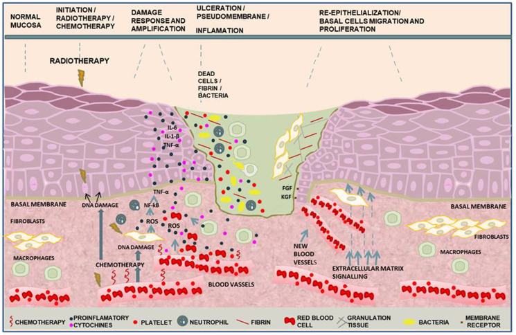
Clinical guidelines for oral mucositis (OM) still consist in palliative care. Herein, we summarize cellular and molecular mechanisms of OM ulceration in response to chemical therapies in animal models. We discuss evidenced anti-inflammatory and anti-oxidant drugs which have not been ever used for OM, such as synthetic peptides as well as cell therapy with mesenchymal stem cells; amniotic membranes, mucoadhesive polymers loaded with anti-inflammatory agents and natural or synthetic electrospun. These approaches have been promising to allow the production of drug-loaded membranes, scaffolds for cells encapsulation or guided tissue regeneration.

Keywords: inflammation; oral mucositis; polymers; regeneration; tissue engineering.


Conflict of interest statement

CONFLICTS OF INTEREST Authors have no conflicts of interest to declare.

Figures

Figure 1
Figure 1. Molecular pathways in the phases of oral mucositis induced by cancer therapy.
In the initiation phase, DNA damage in the basal epithelial cells and mesenchymal cells induced by anticancer treatment leads to reactive oxygen species (ROS) production and cell death. Injury response and amplification induced during radiotherapy and/or chemotherapy by necrotic molecules activate nuclear factor-κβ (NF-κβ) that regulates genes to IL-1-β, IL-6 and tumor necrosis factor α (TNF-α) pro-inflammatory cytokines that damage endothelial cells and fibroblasts. Endothelial membrane breakdown leads to extravasation of red blood cells and platelets to the site of injury resulting in clot formation. The recruitment of neutrophils and macrophages to the site of injury establishes the inflammatory process. Oral cavity bacteria and funghi colonize the clot containing dead cells onto the established ulceration. Bacteria cell wall products recruit macrophages towards the site of injury amplifying the damage. As soon as ROS production ceases, re-epithelialization occurs. Mesenchymal-epithelial cells interaction leads to growth factors release. Vascular endothelial growth factor (VEGF), platelet derived growth factor (PDGF), transforming growth factor (TGF-α), fibroblast growth factor (FGF) and keratinocyte growth factor (KGF), collectively induce new blood vessels formation, keratinocytes proliferation and epithelial cells differentiation.
Figure 2
Figure 2. Evolution of human OM during chemoradiotherapy.
(A) Initial aspect of lower labial mucosa without any change; (B) Mucosa erythema; (C) Initial break in the mucosa represented by a discrete ulceration covered by a pseudomembrane; (D) Confluent ulcerations covered by a fibrinous pseudomembrane; and (E) Areas of necrosis. Informed consent was obtained from the patients to publish images related to diagnosis of oral mucositis.
Figure 3
Figure 3. Electrospun PLGA scaffold for tissue engineering application.
(A) Randomly electrospun microfibers in the format of membrane or film, created by Dr. Raquel Pires Gonçalves and Dr. Marcos Dias Lopes at the Institute of Macromolecules Professor Eloisa Mano of the Federal University of Rio de Janeiro. Scale bar: 4.5 cm; (B) Scanning electron microscopy image of PLGA membrane showing randomly oriented microfibers. Scale bar: 2.5 μm; (CD) Schematic representation of the polymeric membranes with adhered cells, drugs or proteins to be carried out to specific biological sites. Scale bar: 2.5 μm. Dr. Marcos Farina obtained the images using the scanning electron microscope (FEI Quanta 250 at 15KV, 2048 x 2048 pixels) at Federal University of Rio de Janeiro.

Similar articles

Cited by

References

    1. Parkhill AL. Oral Mucositis and Stomatitis Associated with Conventional and Targeted Anticancer Therapy. Journal of Pharmacovigilance. 2013; 1:112.
    1. Treister N, Sonis S. Mucositis: biology and management. Curr Opin Otolaryngol Head Neck Surg. 2007; 15:123–29. 10.1097/MOO.0b013e3280523ad6. - DOI - PubMed
    1. Bowen JM, Gibson RJ, Keefe DM. Animal models of mucositis: implications for therapy. J Support Oncol. 2011; 9:161–68. 10.1016/j.suponc.2011.04.009. - DOI - PubMed
    1. Newton K, Dixit VM. Signaling in innate immunity and inflammation. Cold Spring Harb Perspect Biol. 2012; 4:a006049. 10.1101/cshperspect.a006049. - DOI - PMC - PubMed
    1. Sonis ST, Elting LS, Keefe D, Peterson DE, Schubert M, Hauer-Jensen M, Bekele BN, Raber-Durlacher J, Donnelly JP, Rubenstein EB, and Mucositis Study Section of the Multinational Association for Supportive Care in Cancer, and International Society for Oral Oncology. Perspectives on cancer therapy-induced mucosal injury: pathogenesis, measurement, epidemiology, and consequences for patients. Cancer. 2004; 100:1995–2025. 10.1002/cncr.20162. - DOI - PubMed

LinkOut - more resources