Review
. 2021 Sep;10(5):585-604.
doi: 10.21037/acs-2021-tviv-80.

Current state of transcatheter mitral valve implantation in bioprosthetic mitral valve and in mitral ring as a treatment approach for failed mitral prosthesis

Affiliations
Review

Current state of transcatheter mitral valve implantation in bioprosthetic mitral valve and in mitral ring as a treatment approach for failed mitral prosthesis

Vratika Agarwal et al. Ann Cardiothorac Surg. 2021 Sep.

Abstract

With heightened awareness of mitral valve disease and improvement in surgical techniques, the use of mitral valve bioprostheses has increased. There is a large aging population with prior surgical valvular interventions. Limited durability of the prosthesis due to valvular degeneration over time may necessitate the need for repair or replacement of the prior prosthesis in the future. This usually entails another surgical intervention in this population with elevated risk for a reoperation. There is an ongoing clinical need for newer, less invasive options that are feasible and carry a lower complication rate. The advent of transcatheter heart valve (THV) therapies has opened up a wide range of therapeutic options for treatment of a failed bioprosthesis. Their safety and feasibility are now well established. This article serves as a review of the currently available THVs for implantation in the mitral position, the pre-procedural assessment, the challenges associated with implantation, as well as outcomes associated with a mitral valve-in-valve (VIV) and a mitral valve-in-ring (VIR) procedure.

Keywords: Valve-in-valve (VIV); mitral valve bioprosthesis; surgical heart valve; transcatheter heart valve (THV); transcatheter mitral valve replacement (TMVR); valve-in-ring (VIR).


Conflict of interest statement

Conflicts of Interest: The authors have no conflicts of interest to declare.

Figures

Figure 1
Figure 1
(A) Three-dimensional imaging of the mitral valve displaying the mitral valve en face in surgeon’s view. (B-E) Multiplanar reconstruction of the mitral valve at the level of the mitral valve annulus in mid diastole where the commissure to commissure measurement as well as the septolateral or the anterior-posterior measurement is done. AV, aortic valve; CC, commissure to commissure distance; SL, septolateral distance; IT, intertrigonal distance; ALC, anterolateral commissure; PMC, posteromedial commissure.
Figure 2
Figure 2
(A) Image depicting the true stent internal dimension and the manufacturer provided stent dimension. (B) Image showing the True ID and Stent ID for a pericardial valve with relatively larger true ID. (C) Image showing the True ID and Stent ID for a porcine prosthesis with the leaflets mounted within the stent frame. ID, internal dimension.
Figure 3
Figure 3
(A) Edwards Sapien Valve; (B) Edwards Sapien XT; (C) Edwards Sapien S3; (D) Inovare Valve; (E) Myval Valve; (F) Lotus Valve; (G) Direct Flow Valve.
Figure 4
Figure 4
Example of a mitral VIV for a failed 29 mm CE SAV. (A) Mid-esophageal TEE view of the mitral valve in 2-chamber demonstrating severe eccentric intravalvular regurgitation. (B) Two-dimensional image demonstrating flail posteromedial leaflet (arrow). (C) 3D MPR with measurement of the internal dimension at the time of the procedure which demonstrated pannus ingrowth and dimensions ranging from 22–24 mm. (D,E) Three-dimensional en face view of the mitral valve after deployment of a 26 mm Sapien S3 Ultra valve. (F) Fluoroscopic image of the valve after deployment. The valve was deployed after aligning the ventricular portion of S3 and CE. VIV, valve-in-valve; TEE, transesophageal echocardiogram; CE, Carpentier-Edwards; SAV, Supra-Annular Valve; MPR, multiplanar reconstruction.
Figure 5
Figure 5
Evaluation for VIR using 3Mensio (Pie Medical Imaging, The Netherlands) CT scan analysis for a failed 30 mm Edwards Physio II complete ring (A,B). Note the pronounced saddle shape of the ring (C,D). Image (E) demonstrates the ideal position of the proposed THV, a 29 mm Sapien S3. The inflow of the THV would be 10.7 mm away from the LVOT, suggesting the neo-LVOT should be sufficiently large. Image (F) shows the neo-LVOT in cross section at its smallest area, measuring 426 mm2, well above the cutoff of 120 mm2. Thus, the risk of outflow tract obstruction is low. VIR, valve-in-ring; THV, transcatheter heart valve; LVOT, left ventricular outflow tract.
Figure 6
Figure 6
Preprocedural assessment cardiac CT in a patient with a failed 29 mm CE SAV. The internal diameter of the valve is 25 mm at the level of the annulus (A). A 26 mm Sapien S3 virtual valve was placed using 3Mensio (Pie Medical Imaging) which demonstrates 6.8 mm of clearance from the interventricular septum (B) and a neo-LVOT area of 312 mm2 (C). A deployment angle (RAO 37°, Caudal 0°) is determined by aligning the inflow of the SAV (D), as well as the aorto-mitral angle (114°) (E). CE, Carpentier-Edwards; SAV, Supra-Annular Valve; LVOT, left ventricular outflow tract.
Figure 7
Figure 7
Transseptal planning using 3Mensio analysis of CT scan. A virtual puncture site can be identified that is central on the fossa. In the AP projection (A), the site should line up with the lowest portion of the SAV inflow, and ideally no more superior than the mid portion of the SAV. The coplanar angle can be used to determine the height of the puncture site above the valve annulus (B). A height of 2.5–3.5 cm is desirable, and more height can be gained by puncturing more posteriorly. Two-dimensional imaging can also be used to visualize the puncture site (C,D). There was a large degree of valve pannus formation in this valve (D). SAV, Supra-Annular Valve.
Figure 8
Figure 8
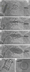
Deployment sequence of a 26 mm Sapien S3 in a 29 mm CE SAV. The mechanism of failure of the SAV was prolapse of two cusps due to leaflet degeneration resulting in severe MR. After delivery of the Confida guidewire, the septal puncture site (yellow arrows) was dilated with a 14×40 mm balloon (A). The valve is delivered into position (B) and the pusher is withdrawn more proximally than typical [*] to increase flexibility of the system during deployment. As the THV balloon is inflated, the valve self-centers as it anchors on the SAV facilitated by the operator in position one applying forward pressure on the system (C,D). Coplanar and en face images of the VIV are shown in images (E,F). (Note: patient also has a Micra leadless pacemaker in the apex of the right ventricle). CE, Carpentier-Edwards; SAV, Supra-Annular Valve; VIV, valve-in-valve; THV, transcatheter heart valve.
Video
Video
Current state of transcatheter mitral valve implantation in bioprosthetic mitral valve and in mitral ring as a treatment approach for failed mitral prosthesis.

References

    1. Virani SS, Alonso A, Benjamin EJ, et al. Heart Disease and Stroke Statistics-2020 Update: A Report From the American Heart Association. Circulation 2020;141:e139-596. 10.1161/CIR.0000000000000757 - DOI - PubMed
    1. Iung B, Baron G, Butchart EG, et al. A prospective survey of patients with valvular heart disease in Europe: The Euro Heart Survey on Valvular Heart Disease. Eur Heart J 2003;24:1231-43. 10.1016/S0195-668X(03)00201-X - DOI - PubMed
    1. Nkomo VT, Gardin JM, Skelton TN, et al. Burden of valvular heart diseases: a population-based study. Lancet 2006;368:1005-11. 10.1016/S0140-6736(06)69208-8 - DOI - PubMed
    1. Dziadzko V, Clavel MA, Dziadzko M, et al. Outcome and undertreatment of mitral regurgitation: a community cohort study. Lancet 2018;391:960-9. 10.1016/S0140-6736(18)30473-2 - DOI - PMC - PubMed
    1. Chatterjee S, Rankin JS, Gammie JS, et al. Isolated mitral valve surgery risk in 77,836 patients from the Society of Thoracic Surgeons database. Ann Thorac Surg 2013;96:1587-94; discussion 1594-5. 10.1016/j.athoracsur.2013.06.051 - DOI - PubMed