. 2021 Oct:10:e00204.
doi: 10.1016/j.ohx.2021.e00204. Epub 2021 May 20.

A low-cost, wearable, do-it-yourself functional near-infrared spectroscopy (DIY-fNIRS) headband

Affiliations

A low-cost, wearable, do-it-yourself functional near-infrared spectroscopy (DIY-fNIRS) headband

Francis Tsow et al. HardwareX. 2021 Oct.

Abstract

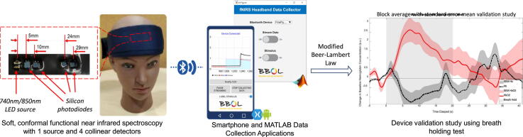
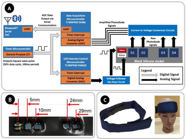
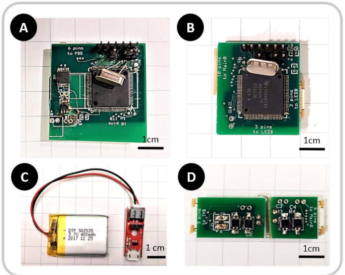
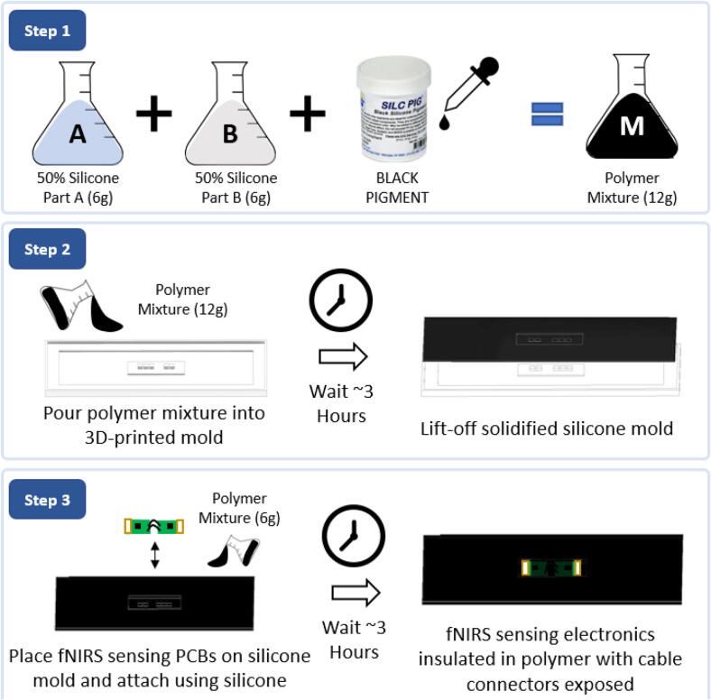
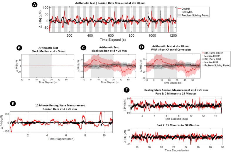
Neuromonitoring in naturalistic environments is of increasing interest for a variety of research fields including psychology, economics, and productivity. Among functional neuromonitoring modalities, functional near-infrared spectroscopy (fNIRS) is well regarded for its potential for miniaturization, good spatial and temporal resolutions, and resilience to motion artifacts. Historically, the large size and high cost of fNIRS systems have precluded widespread adoption of the technology. In this article, we describe the first open source, fully integrated wireless fNIRS headband system with a single LED-pair source and four detectors. With ease of operation and comfort in mind, the system is encased in a soft, lightweight cloth and silicone enclosure. Accompanying computer and smartphone data collection software have also been provided, and the hardware has been validated using classic fNIRS tasks. This wear-and-go design can easily be scaled to accommodate a larger number of fNIRS channels and opens the door to easily collecting fNIRS data during routine activities in naturalistic conditions.

Keywords: brain imaging; functional near infrared spectroscopy; hemodynamics; neuroimaging.


Conflict of interest statement

The authors declare that they have no known competing financial interests or personal relationships that could have appeared to influence the work reported in this paper.

Figures

None
Graphical abstract
Fig. 1
Fig. 1
A) Block diagram illustrating the interconnectivity of the various subcomponents constituting the fNIRS sensing control circuitry. B) Photograph (not to scale) of a prototype optode. C) Photograph showing a model wearing our fNIRS headband device.
Fig. 2
Fig. 2
Images of assembled control circuits. A) Data acquisition and Bluetooth® broadcasting board, B) LED Intensity control board, C) Detachable Sparkfun® LiPo Charger with 400 mAh LiPo battery, D) fNIRS sensing electronics.
Fig. 3
Fig. 3
Silicone polymer skin assembly for insulating and protecting the fNIRS sensing electronics. Step 1) Prepare polymer mixture by mixing equal parts of silicone parts A and B with 6 drops of black pigment. Step 2) Pour silicone and set polymer mold for three hours, then lift-off. Step 3) Place fNIRS sensing electronics on lifted-off silicone mold and set in place using a fresh batch of polymer mixture.
Fig. 4
Fig. 4
Cloth enclosure assembly. Step 1) Laser-cut cloth material using the provided “clothLaserCut” file in the OSF repository. Step 2) Sew on Velcro® hook and loop sides to designated areas to form an enclosable headband. Step 3) Fold cloth enclosure to form a “tube” (held together with Velcro®) to be wrapped around the head of a user.
Fig. 5
Fig. 5
Consolidation of the remaining components of the fNIRS headband system. Step 1) Apply a small amount of silicone polymer (2 g each of Part A and B) along the tapered edges of the silicone polymer encasing the fNIRS electronics and let dry for three hours. Step 2) Connect flat 6-pin and 10-pin cables as demonstrated in the diagram above to complete assembly of the system. The system can be powered by plugging in the LiPo Charger using the provided female headers.
Fig. 6
Fig. 6
A) Screenshot of the Android phone application developed using Xamarin.Android. The application allows users to collect raw voltage readings from the fNIRS headband system on the phone for access later. During data collection, users can check the device connection status and annotate data when “stimuli” are applied. B) Screenshot of the MATLAB® computer application to easily connect to and save data from the fNIRS headband system.
Fig. 7
Fig. 7
Physiological validation using a breath-holding test. A) Complete session data. The solid red lines represent OxyHb, dashed black lines are DeoxyHb, and shaded gray areas represents breath-holding periods. Characteristic dips and recovery in OxyHb are visible in these data. B thru E) Block median with highlighted standard error of the median of 10 cycles measuring the change in baseline measurement with respect to the 10 s preceding the breath-holding period. An increase in source-detector separation results in deeper imaging into the head F) A segment of raw, unprocessed data from our fNIRS device during 850-nm LED light input is shown. Arterial pulsations are visible in the figure. (For interpretation of the references to color in this figure legend, the reader is referred to the web version of this article.)
Fig. 8
Fig. 8
A) Complete session data of Arithmetic Test. The solid red lines represent OxyHb, dashed black lines DeoxyHb, and shaded gray area represent problem solving periods. Characteristic rises of OxyHb during problem solving phases are visible. B thru D) Block median with highlighted standard error median of 20 cycles measuring change in baseline measurement with respect to the 10 s preceding arithmetic problem-solving period. In B and C, change in hemoglobin concentrations are shown from the shortest detector at d = 5 mm and the farthest detector d = 28 mm. In D, the short channel signal from d = 5 mm is subtracted from long channel signal at d = 28 mm to demonstrate short channel correction. E) Resting state fNIRS measurements for 10 min. F) Resting state fNIRS measurements for 30 min. Both data demonstrate stability of the measurements throughout the imaging durations. (For interpretation of the references to color in this figure legend, the reader is referred to the web version of this article.)

References

    1. Pinti P., Aichelburg C., Gilbert S., Hamilton A., Hirsch J., Burgess P., Tachtsidis I. A review on the use of wearable functional near-infrared spectroscopy in naturalistic environments. Jpn. Psychol. Res. 2018;60(4):347–373. doi: 10.1111/jpr.12206. - DOI - PMC - PubMed
    1. A. Hamilton, P. Pinti, D. Paoletti, J.A. Ward, Seeing into the Brain of an Actor with Mocap and fNIRS, in: ISWC’18 Proc. 2018 ACM Int. Symp. Wearable Comput., 2018: pp. 216–217. DOI:10.1145/3267242.3267284.
    1. T.(舟根司) Funane, H.(敦森洋和) Atsumori, A.(木敦) Suzuki, M.(木口雅史) Kiguchi, Noncontact brain activity measurement system based on near-infrared spectroscopy, Appl. Phys. Lett. 96 (2010) 123701. DOI:10.1063/1.3367737.
    1. McKendrick R., Mehta R., Ayaz H., Scheldrup M., Parasuraman R. Prefrontal hemodynamics of physical activity and environmental complexity during cognitive work. Hum. Factors. 2017;59(1):147–162. doi: 10.1177/0018720816675053. - DOI - PubMed
    1. K. Izzetoglu, S. Bunce, M. Izzetoglu, B. Onaral, K. Pourrezaei, Functional near-infrared neuroimaging, in: Proc. 26th Annu. Int. Conf. IEEE Eng. Med. Biol. Soc. Vols 1-7, 2004: pp. 5333–5336. - PubMed

LinkOut - more resources