Review
. 2022 Jan;480(1):65-84.
doi: 10.1007/s00428-021-03182-7. Epub 2021 Nov 3.

Papillary lesions of the breast

Affiliations
Review

Papillary lesions of the breast

Janina Kulka et al. Virchows Arch. 2022 Jan.

Abstract

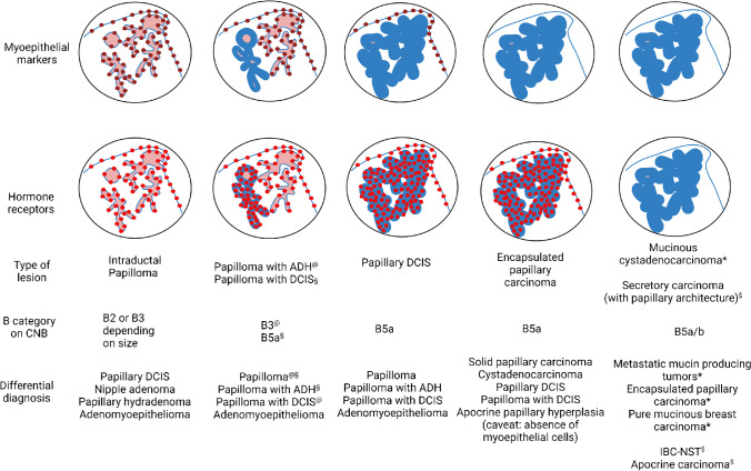
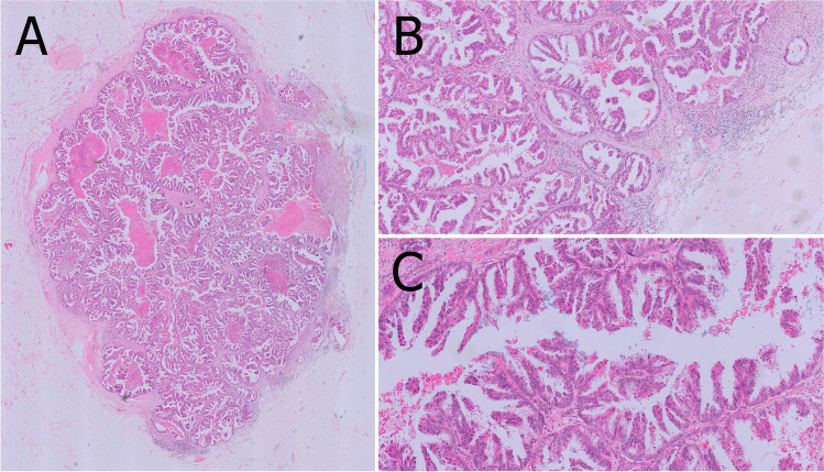
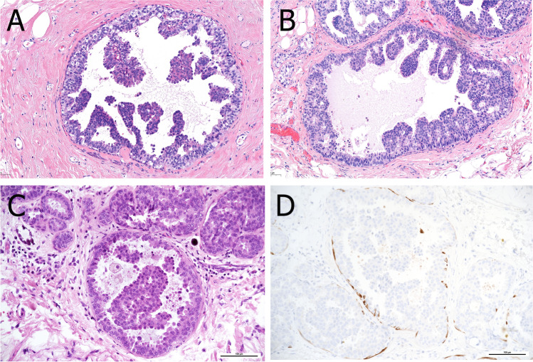
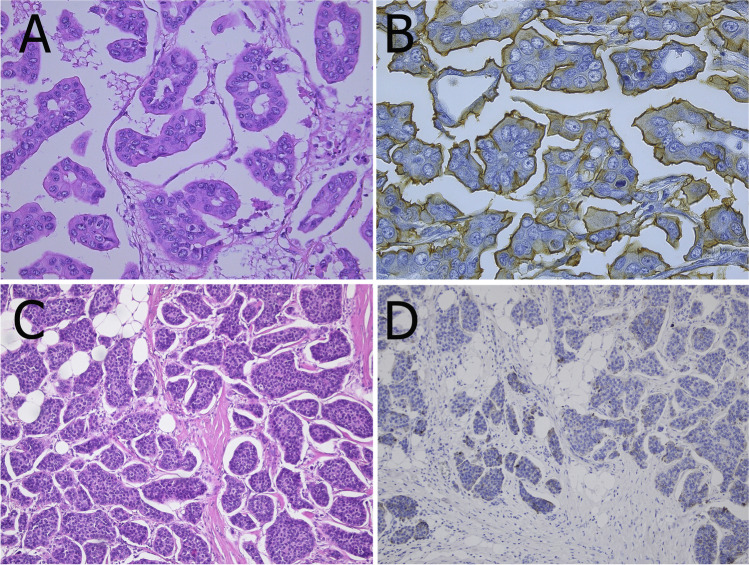
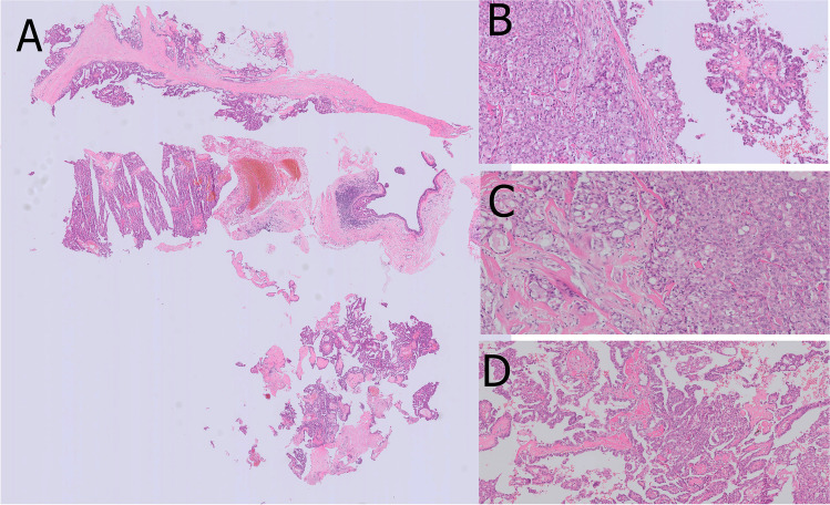
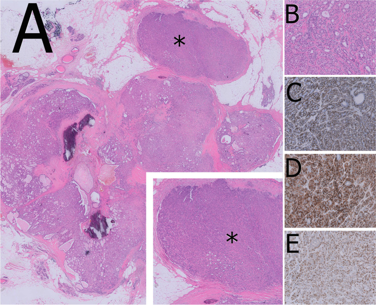
Papillary lesions of the breast represent a heterogeneous group of lesions including benign papillomas, papillomas with focal epithelial atypia, fully fledged ductal carcinoma in situ (DCIS) or lobular neoplasia, papillary DCIS, encapsulated papillary carcinomas without or with invasion, solid papillary carcinomas, and invasive papillary carcinomas. A micropapillary pattern characterized by lack of fibrous stalks within the papillae is observed in micropapillary DCIS and invasive micropapillary carcinoma. In addition, a variety of other rare breast lesions reveals a papillary architecture such as tall cell carcinoma with reversed polarity (TCCRP) and mucinous cystadenocarcinoma, adenomyoepithelioma, and secretory carcinoma. In addition, benign lesions such as usual ductal hyperplasia, apocrine metaplasia, gynecomastia, and juvenile papillomatosis may show a papillary or micropapillary architecture. Fragments of a benign papilloma in a breast biopsy are considered a lesion of uncertain malignant potential (B3 in the European classification) and excision is mostly recommended. Although the knowledge about molecular pathology of papillary breast lesions has increased, there is not sufficient evidence for diagnostically useful molecular features, yet. The aim of this review is to provide an update on papillary and micropapillary lesions with emphasis on problematic areas for daily diagnostic work including biopsies.

Keywords: Biopsy; Breast; DCIS; Ductal carcinoma in situ; Micropapillary; Papillary lesions.


Conflict of interest statement

Dr. Lax reports personal fees from Roche, AstraZeneca, Novartis, and Biogena outside the submitted work. All other authors declare no conflict of interest.

Figures

Fig. 1
Fig. 1
Staining patterns of myoepithelial markers and hormone receptors in various papillary lesions of the breast. The presence of myoepithelial markers is illustrated by bordeaux brown dots. Myoepithelial cells may be present or not in the peripheral wall and/or centrally in association with the branching fibrovascular cores illustrated in pink. In the same fashion, the bright red dots highlight the expression of hormone receptors in the lining epithelium. The lining epithelium is illustrated with a continuous blue line which depending on the degree of proliferation, and malignancy of the lesion becomes thicker with almost disappearance of the pink fibrovascular cores. Each combination of staining patterns is associated to specific lesions for which the B category on CNB, and the differential diagnosis is proposed. (This figure was created with BioRender.com)
Fig. 2
Fig. 2
Papilloma in a large duct (central location) showing sclerotic areas (A and B). A prominent myoepithelial cell layer highlights the typical “two cell types” of benign papillary lesions (C). Myoepithelial cells can be demonstrated by p63 immunoreactivity both at the peripheral wall and along the fibrovascular cores (D)
Fig. 3
Fig. 3
Sclerosis of the papilloma’s capsule with entrapped epithelial and myoepithelial cells may mimic a pseudo-infiltrative pattern (A). Myoepithelial markers such as p63 demonstrate the presence of myoepithelial cells and exclude invasion (B)
Fig. 4
Fig. 4
Intraductal papilloma with atypical epithelial proliferation of ductal type in an area of < 3 mm size qualifying for the diagnosis of ADH (A). The atypical epithelial proliferation lacks CK5 immunoreactivity (B). Intraductal papilloma with lobular neoplasia characterized by a solid proliferation of monomorphic cells with reduced cell cohesion (C, D). The lobular neoplasia is characterized by lack of immunoreactivity for CK5 (E) and e-cadherin (F)
Fig. 5
Fig. 5
Papillary DCIS (A) with microinvasion (*). Cribriform architecture is present at the periphery of involved ducts (B). Characteristic features are low-grade nuclei (C), lack of myoepithelial cells as demonstrated by p63 (D) and diffuse, strong ER positivity (E)
Fig. 6
Fig. 6
Encapsulated papillary carcinoma with typical gross appearance (A). An arborizing papillary structure lined by low-grade atypical epithelium is present within a cystically dilated space surrounded by a fibrotic capsule (B) and may be associated with frank invasion (C)
Fig. 7
Fig. 7
Solid papillary carcinoma characterized by a low-grade atypical epithelial proliferation with solid growth pattern filling dilated ducts (A). Invasion is characterized by small infiltrative ducts and nests (B). Neuroendocrine differentiation is frequently found as confirmed by expression of synaptophysin (C)
Fig. 8
Fig. 8
Invasive papillary carcinoma characterized by cystically dilated spaces of various size (A) and frank invasion with focal necrosis at the periphery (B). Delicate arborizing papillary structures are readily appreciated at medium power magnification (C)
Fig. 9
Fig. 9
TCCRP showing multinodular architecture with presence of fibrovascular cores centrally located in the nodular structures (A). Narrow fibrous septae (B) and occasionally colloid-like eosinophilic material (C) are characteristic features
Fig. 10
Fig. 10
Mucinous cystadenocarcinoma consisting of cysts filled with abundant mucin (A). The delicate fibrous septa are covered by eosinophilic tumor cells with floating cellular morules on top (C). The tumor cells may show abundant mucin production (D)
Fig. 11
Fig. 11
Micropapillary and papillary DCIS in a small duct (A) adjacent to invasive papillary carcinoma (not shown). Pure micropapillary DCIS is characterized by micropapillary projections lacking fibrovascular cores and typical triangular architecture observed in micropapillary hyperplasia (B, C). Immunohistochemistry for CK5 shows the lack of basally differentiated cells (D)
Fig. 12
Fig. 12
Invasive micropapillary carcinoma characterized by an “inside-out” growth pattern (A) highlighted by EMA immunoreactivity (B). In contrast, EMA is negative in invasive NST carcinomas with retraction clefts (C, D)
Fig. 13
Fig. 13
Secretory carcinoma with papillary architecture in a core needle biopsy (A). Solid areas (B) in transition to microcystic areas with presence of intraluminal eosinophilic secretion (C) and prominent papillary architecture (D) are typical cytoarchitectural features
Fig. 14
Fig. 14
Adenomyoepithelioma with partial papillary growth pattern showing multinodular architecture with thick fibrovascular septa (A) and presence of expansive nodules of myoepithelial cells (*). The myoepithelial nodules show a mixture of glandular adenosis-like and spindle cell growth patterns (B) Proliferation of myoepithelial cells is confirmed by CK14 (C), calponin (D), and p63 immunostaining (E)
Fig. 15
Fig. 15
Intraductal papilloma with myoepithelial hyperplasia showing an area with increased cellularity (asterisk) (A) characterized by expansion of myoepithelial cells (B). The retained myoepithelial layer and the expansion of the myoepithelial compartment is highlighted by p63 (C) and CD10 (D) immunoreactivity
Fig. 16
Fig. 16
Nipple adenoma presenting as a mass at the dermo-epidermal junction with a large duct at the periphery (A). Focally, usual ductal hyperplasia with papillary architecture is evident (B)

References

    1. Abdulwaasey M, Tariq MU, Minhas K, Kayani N. Invasive breast carcinoma arising in a nipple adenoma after 15 years: report of a rare case and literature review. Cureus. 2020;12:e8586. doi: 10.7759/cureus.8586. - DOI - PMC - PubMed
    1. Acs G, Khakpour N, Kiluk J, Lee MC, Laronga C. The presence of extensive retraction clefts in invasive breast carcinomas correlates with lymphatic invasion and nodal metastasis and predicts poor outcome: a prospective validation study of 2742 consecutive cases. Am J Surg Pathol. 2015;39:325–337. doi: 10.1097/PAS.0000000000000339. - DOI - PubMed
    1. Adrada B, Arribas E, Gilcrease M, Yang WT. Invasive micropapillary carcinoma of the breast: mammographic, sonographic, and MRI features. AJR Am J Roentgenol. 2009;193:W58–63. doi: 10.2214/AJR.08.1537. - DOI - PubMed
    1. Agoumi M, Giambattista J, Hayes MM. Practical considerations in breast papillary lesions: a review of the literature. Arch Pathol Lab Med. 2016;140:770–790. doi: 10.5858/arpa.2015-0525-RA. - DOI - PubMed
    1. Alsadoun N, MacGrogan G, Truntzer C, Lacroix-Triki M, Bedgedjian I, Koeb MH, El Alam E, Medioni D, Parent M, Wuithier P, Robert I, Boidot R, Arnould L. Solid papillary carcinoma with reverse polarity of the breast harbors specific morphologic, immunohistochemical and molecular profile in comparison with other benign or malignant papillary lesions of the breast: a comparative study of 9 additional cases. Mod Pathol. 2018;31:1367–1380. doi: 10.1038/s41379-018-0047-1. - DOI - PubMed

MeSH terms