

doi: 10.1164/rccm.202103-0594IM.
The Bronchial Circulation in COVID-19 Pneumonia
Affiliations
- PMID: 34734553
- PMCID: PMC8865596
- DOI: 10.1164/rccm.202103-0594IM
Item in Clipboard
The Bronchial Circulation in COVID-19 Pneumonia
Am J Respir Crit Care Med.
.
No abstract available
Figures
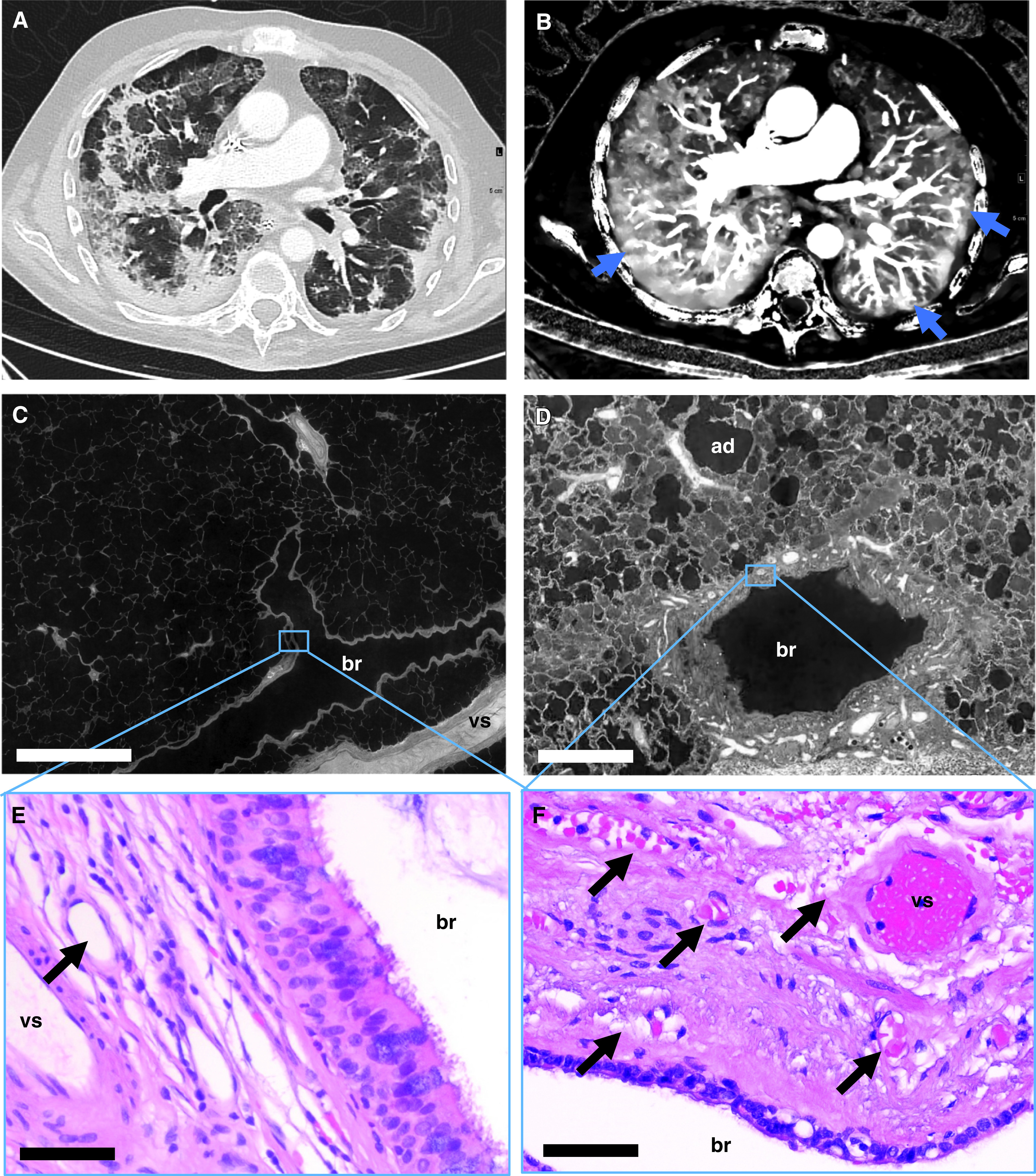
Bronchial artery and vasa vasorum dilatation and expansion in coronavirus disease (COVID-19) pneumonia. (A) Dual energy computed tomography revealed the characteristic features of severe COVID-19 pneumonia as bilateral ground-glass opacities, peribronchial consolidations, and a diffuse crazy-paving pattern. (B) Iodine map showed dilatation of macrovessels in or leading to opacified air spaces and frequently evident vascular “tree-in-bud” (blue arrows). (C and D) Hierarchical phase-contrast tomography (high-resolution tomographic images analyzed by Synchrotron radiation X-ray tomographic microscopy) demonstrated the regular architecture in healthy control lungs (C), whereas in COVID-19 pneumonia, a dilatation and an expansion of vasa vasorum and peribronchial vessels was observed (D). Scale bars: C, 1,000 μm; D, 400 μm. (E and F) Representative hematoxylin and eosin–stained sections show sporadic peribronchial vessels (black arrow) in the control tissue (E), whereas the peribronchial plexus in COVID-19 lungs was characterized by an expansion and the formation of angiomatoid- and plexiform-like vessels (black arrows) (F). Scale bars: 20 μm. ad = alveolar duct; br = bronchus; vs = pulmonary vessel.
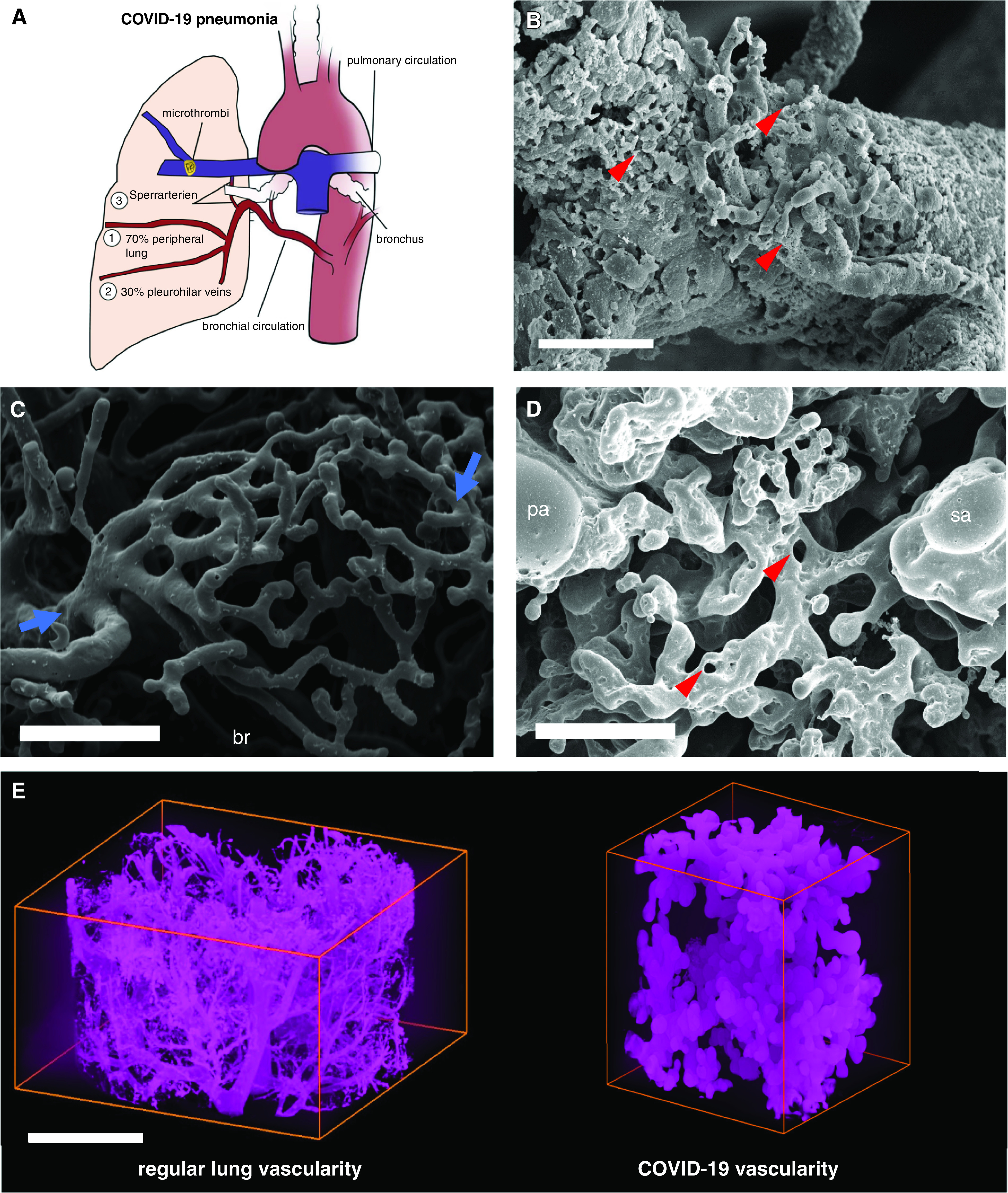
Intrapulmonary shunting via bronchial circulation in coronavirus disease (COVID-19) pneumonia. (A) The schematic illustrates intrapulmonary cross-circulation and draining by bronchial arteries to the peripheral lung, pleurohilar veins, and “Sperrarterien,” specialized, spiral-like intrapulmonary arteriovenous anastomoses recruited by hypoxia and flow irregularities. The bronchial circulation arises primarily direct from the thoracic aorta and intercostal arteries and, under normal conditions, constitutes less than 2% volume of the left cardiac output. However, the bronchial flow can significantly exceed the regular hemodynamics upon hypoxic and inflammatory stimulus. As part of a physiological right–left shunting, nearly two-thirds of the bronchial vessels are drained peribronchially to the peripheral lung and finally into the left ventricle (up to 4% of left ventricular output), whereas the remaining third is perfused pleurohilar veins draining ultimately to the right heart. (B) Scanning electron micrograph of a microvascular corrosion cast demonstrates the expansion of vasa vasorum of an intralobular artery by intussusceptive angiogenesis (red arrowheads) in the lung tissue of a patient who succumbed to severe COVID-19 pneumonia. Scale bar, 50 μm. Intussusceptive angiogenesis is a well-characterized morphogenetic process observed during the capillary expansion in cancer, inflammation, and regeneration, but it has also been reported in the alveolar plexus of COVID-19 lungs. Scale bar, 100 μm. (C) Scanning electron micrograph of a microvascular corrosion cast demonstrates the peribronchial plexus in healthy control lungs (blue arrows). Scale bar, 200 μm. (D) Scanning electron micrograph of a microvascular corrosion cast reveals the cross-circulation in COVID-19 lung tissue between a Sperrarterie and a lobular artery in a secondary pulmonary lobule, which is characterized by plexiform-like vascular proliferations by intussusceptive angiogenesis (red arrowheads). Scale bar, 200 μm. (E) Three-dimensional evaluation of microvascular corrosion casts by Synchrotron radiation X-ray tomographic microscopy illustrating the irregular, hierarchical vascularity with dilated, blunt-like vessels in COVID-19 pneumonia. Scale bar, 100 μm. br = bronchiole, pa = lobular artery; sa = Sperrarterie.
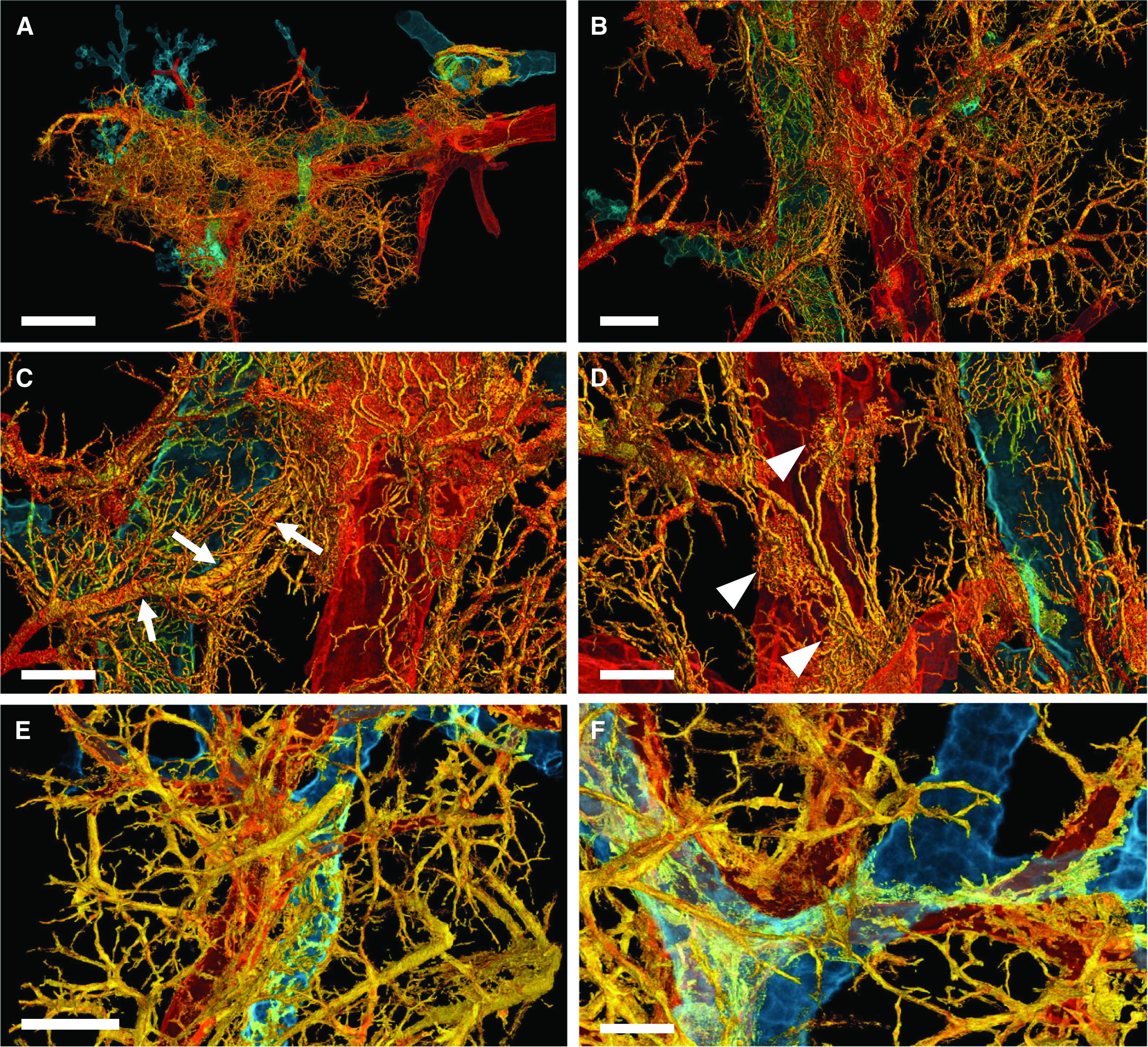
Three-dimensional complexity of vascular remodeling and bronchiopulmonary shunting in coronavirus disease (COVID-19) highlighted by hierarchical phase-contrast tomography. (A) The distal bronchiole (blue) is paralleled by a centrilobular pulmonary artery (red) and an expanded plexus of private vessels (vasa vasorum) (yellow) in a secondary pulmonary lobule in the periphery of the left lower lobe. Scale bar, 4 mm. (B) Dilatation and expansion of the peribronchiolar plexus and vasa vasorum (yellow) is observed around the distal terminal bronchiole (blue) and centrilobular pulmonary artery (red). Scale bar, 1.3 mm. (C) Numerous arteriovenous anastomoses (yellow) form between bronchial arteries and pulmonary artery drain into a pulmonary vein accompanied by a pronounced shunting of a Sperrarterie (white arrows). These specialized arteries are essential for intrapulmonary arteriovenous shunting and are located mainly at the septal margin of secondary pulmonary lobules, where up to 3 to 5 such “Sperrarterien” anastomoses can be found in each lobule. Scale bar, 0.7 mm. (D) The perivascular vasa vasorum and bronchial vascular plexus are characterized by complex nodular lesions with angiomatoid- and plexiform-like arrangements (white arrowheads). Scale bar, 0.7 mm. For high-resolution details, see COVID-19 Pneumonia in the online supplement . (E) In controls, the vascular architecture shows a well-organized hierarchy of peribronchial vessels. Scale bar, 1.3 mm. (F) Only few perivascular vasa vasorum are observed in healthy control tissue. Scale bar, 0.7 mm. For high-resolution details, see Control in the online supplement.
Comment in
-
Bronchial Mucosal Microcirculation in SARS-CoV-2 Infection: Role in Innate Humoral Defense?Am J Respir Crit Care Med. 2022 Jul 15;206(2):228-229. doi: 10.1164/rccm.202202-0412LE. Am J Respir Crit Care Med. 2022. PMID: 35533389 Free PMC article. No abstract available.
-
Microvascular Shunts in COVID-19 Pneumonia.Am J Respir Crit Care Med. 2022 Jul 15;206(2):227-228. doi: 10.1164/rccm.202111-2627LE. Am J Respir Crit Care Med. 2022. PMID: 35533392 Free PMC article. No abstract available.
References
-
- McCullagh A, Rosenthal M, Wanner A, Hurtado A, Padley S, Bush A. The bronchial circulation--worth a closer look: a review of the relationship between the bronchial vasculature and airway inflammation. Pediatr Pulmonol . 2010;45:1–13. - PubMed
Publication types
MeSH terms
Grants and funding
- MR/S007687/1/MRC_/Medical Research Council/United Kingdom
- 2020-225394/Chan Zuckerberg Initiative
- CiET1819/10/Royal Academy of Engineering
- F32 HL134229/HL/NHLBI NIH HHS/United States
- KFO311/Deutsche Forschungsgemeinschaft
- R01 HL094567/HL/NHLBI NIH HHS/United States
- HL94567/NIH and NHLBI
- 771883/H2020 European Research Council Consolidator
- MR/R025673/1/MRC_/Medical Research Council/United Kingdom
- DEFEAT PANDEMIcs, 01KX2021/Bundesministerium für Bildung und Forschung
- XHale/H2020 European Research Council Consolidator
- HL134229/NIH and NHLBI
LinkOut - more resources
Full Text Sources
Medical
