. 2021 Nov 4:10:e69786.
doi: 10.7554/eLife.69786.

A WDR35-dependent coat protein complex transports ciliary membrane cargo vesicles to cilia

Affiliations

A WDR35-dependent coat protein complex transports ciliary membrane cargo vesicles to cilia

Tooba Quidwai et al. Elife. .

Abstract

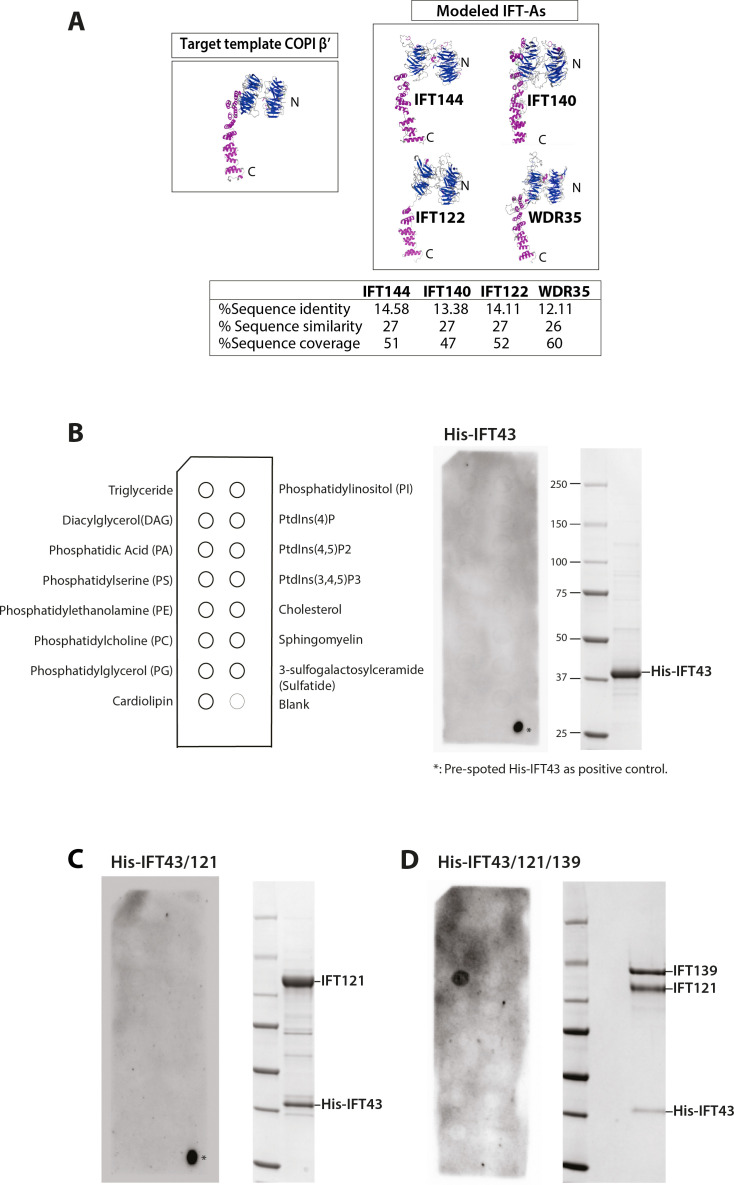
Intraflagellar transport (IFT) is a highly conserved mechanism for motor-driven transport of cargo within cilia, but how this cargo is selectively transported to cilia is unclear. WDR35/IFT121 is a component of the IFT-A complex best known for its role in ciliary retrograde transport. In the absence of WDR35, small mutant cilia form but fail to enrich in diverse classes of ciliary membrane proteins. In Wdr35 mouse mutants, the non-core IFT-A components are degraded and core components accumulate at the ciliary base. We reveal deep sequence homology of WDR35 and other IFT-A subunits to α and ß' COPI coatomer subunits and demonstrate an accumulation of 'coat-less' vesicles that fail to fuse with Wdr35 mutant cilia. We determine that recombinant non-core IFT-As can bind directly to lipids and provide the first in situ evidence of a novel coat function for WDR35, likely with other IFT-A proteins, in delivering ciliary membrane cargo necessary for cilia elongation.

Keywords: CLEM; COPI; IFT; TEM; cell biology; chlamydomonas reinhardtii; cilia; ciliary pocket; coatomer; correlative light and electron microscopy; intraflagellar transport; membrane cargos; mouse; transmission electron microscopy; vesicular traffic.

Plain language summary

Most human cells have at least one small hair-like structure on their surface called a cilium. These structures can act as antennae and allow the cell to sense signals from the rest of the body. To do this, they contain proteins that differ from the rest of the cell. The content of cilia depends on regulated delivery of these proteins in and out of cilia by a process called the intraflagellar transport or IFT, which involves a large complex made of several proteins. This complex shuttles the cargo proteins back and forth between the base and the tip of the cilia. However, ciliary proteins are not produced in the cilia; instead, they are made in a different part of the cell and then they are transported to the ciliary base. At the point where they enter the cilia, they were thought to bind to the assembling IFT ‘trains’ and be transported across the ciliary gate to the positions where they are needed in cilia. One of the components of the IFT machinery is a protein called WDR35, also known as IFT121. If the gene that codes for this protein is faulty or missing, it results in severe disorders in both humans and mice including a range of potentially lethal skeletal dysplasias. Interestingly, without WDR35, cells cannot build functional cilia. The absence of this protein not only disrupts IFT, stopping certain ciliary proteins and their associated membranes from entering cilia; it also causes a ‘traffic jam’ with a pile-up of transport intermediates from the place in cell where they are made to the cilia. It is unclear why a mutation in one of the components of the IFT would have this effect, raising the question of whether WDR35, or IFTs a whole, has another role in bringing the cargo proteins into the cilia. To understand this phenomenon, Quidwai et al. analysed the structure of WDR35 and other IFT proteins and found that they are very similar to a protein complex called COPI, which is involved in transporting membrane proteins around the cell. When certain proteins are newly made, they are stored in small lipid bubbles – called vesicles – that then selectively move to where the proteins are needed. COPI coats these vesicles, helping them get to where they need to go in a process called vesicular transport. Quidwai et al. found that WDR35 and other IFT proteins are able to bind to specific types of lipid molecules, suggesting that they might be assisting in a form of vesicle transport too. Indeed, when mouse cells grown in the lab were genetically engineered so they could not produce WDR35, coatless vesicles accumulated around the base of the cilia. Adding back WDR35 to these mutant cells rescued these defects in vesicle transport to cilia as well as allowed functional cilia to be formed. These results provide evidence that WDR35, likely with other IFT proteins, acts as a COPI-like complex to deliver proteins to growing cilia. Further research will investigate the composition of these vesicles that transport proteins to cilia, and help pinpoint where they originate. Quidwai et al.’s findings not only shed light on how different genetic mutations found in patients with cilia dysfunction affect different steps of transporting proteins to and within cilia. They also increase our understanding of the cellular roadmap by which cells shuttle building blocks around in order to assemble these important ‘antennae’.


Conflict of interest statement

TQ, JW, EH, NP, WL, PK, JW, LM, MK, JM, EL, GP, PM No competing interests declared

Figures

Figure 1.
Figure 1.. Wdr35-/- and Dync2h1-/- mutant cells have a drastic reduction in cilia length but have no difference in the number of cilia.
(A) Wild type (WT) and mutant mouse embryonic fibroblasts (MEFs) and those rescued by transiently expressing WDR35-EmGFP serum-starved for 24 hr, fixed and stained with acetylated α-tubulin (green) and γ-tubulin (magenta), nuclei (blue). Boxed regions are enlarged below, and arrows point at ciliary axoneme stained for acetylated α-tubulin. (B) Quantification of cilia length for acetylated α-tubulin. n = total number of cells from three different biological replicates (represented by different shapes). Asterisk denotes significant p-value from t-test: *p<0.05, **p<0.01, ***p<0.001. (C) Percentage of acetylated α-tubulin-positive ciliated cells. (D) 24 hr serum-starved WT and mutant MEFs stained for nuclei (blue), acetylated α-tubulin/polyglutamylated tubulin (green), rootletin (cyan), and transition zone proteins MKS1/NPHP-1 (magenta) show no difference in the localization of transition zone proteins MKS1 and NPHP-1. Gray scale enlarged regions are labeled green (G), magenta (M), and cyan (C). (E) Schematic of intraflagellar transport (IFT) pathway in cilia.
Figure 1—figure supplement 1.
Figure 1—figure supplement 1.. The organization of centriolar satellites (CS) around Wdr35-/- cilia is not changed.
CS marker PCM1 intensity and localization are unchanged in Wdr35 +/+ and Wdr35-/- mouse embryonic fibroblasts (MEFs) serum-starved for 24 hr to induce ciliogenesis and imaged (A) fixed after staining with antibodies; PCM1 (magenta) and γ-tubulin (green). Nuclei are in blue. (B) Quantification of PCM1 intensity around the centrosome in concentric rings of 1 µm around the basal body (γ-tubulin). n = 50 cells (three biological replicates each). (C) Imaged live after staining with SNAP-TMR dye for endogenous SNAP tagged PCM1 (magenta) and microtubule marker SiR-tubulin (gray). These cells are also expressing ARL13B-EGFP (green) (Figure 1—video 1).
Figure 2.
Figure 2.. Wdr35-/- cilia exhibit retrograde transport defects of IFT-B, similar to Dync2h1-/-, although IFT-B complex assembly is unaffected.
(A) IFT-B (green) accumulates beyond the axoneme (Ac-α-tubulin, magenta) in Wdr35 and Dync2h1 mutant cilia from 24 hr serum-starved and fixed mouse embryonic fibroblasts (MEFs). (B) Length quantification shows IFT-B accumulates beyond acetylated α-tubulin in significantly shorter mutant cilia. n = total number of cells from three different biological replicates represented by different shapes. Asterisk denotes significant p-value from t-test: *p<0.05, **p<0.01, ***p<0.001. Scale bars = 5 µm. (C, D) Despite differences in localization, IFT88 immunoprecipitation/mass spectrometry (IP/MS) analysis of E11.5 wild type (WT) and Wdr35-/- littermate embryos reveals no difference in the composition of the IFT-B complex. Antibody highlights bait (IFT88) for IP. (C) Normalized label-free quantification intensities (LFQs) to IFT88 intensity reveal no difference between WT and Wdr35-/- IFT-B complex composition. N = 4 embryos/genotype. (D) The number of unique peptides identified in IP/MS.
Figure 3.
Figure 3.. WDR35 is essential for the stability and recruitment of the IFT-A complex into cilia.
(A) Immunoprecipitation/mass spectrometry (IP/MS) data show the stability of the IFT-A complex is disrupted in Wdr35-/- lysates. N = 6 embryos/genotype. Antibody highlights bait (IFT140) for IP. (B) Immunoblots confirm the non-core IFT-A complex is unstable in Wdr35 mutants. IFT43 runs close to the molecular weight of IgG and is shown by an arrow as IFT43 band over the IgG band from IFT140 IP in wild type (WT). The corresponding band is absent in Wdr35 null samples. (C, D) Immunoblots for the total level of IFT-A subunits in E11.5 embryo lysates show non-core components IFT139 and IFT43 to be missing in Wdr35 mutants (C), quantified by densitometry (D). N = biological replicates. Asterisk denotes significant p-value from t-test: *p<0.05, **p<0.01, ***p<0.001. (E) Inhibition of the proteasome by treatment with MG-132 rescues IFT43 stability in Wdr35-/- mouse embryonic fibroblasts (MEFs). (F) MEFs serum-starved for 24 hr reveal a retrograde transport defect in Dync2h1-/- versus a failed recruitment of IFT-A proteins into Wdr35-/- cilia. Cells are fixed and stained for respective IFT-A (green) and γ- and acetylated α-tubulin (magenta). Arrowheads point at cilia. Scale bars = 5 µm. Due to a lack of specific immunoreagents, IFT122 signal is from transiently expressed Ift122-GFP. All other panels represent endogenous signal detected by IF.
Figure 3—figure supplement 1.
Figure 3—figure supplement 1.. WDR35 is essential for the stability and recruitment of the IFT-A complex into cilia.
(A, B) Immunoblots for the total level of IFT-A subunits in mouse embryonic fibroblast (MEF) lysates show non-core components IFT139 and IFT43 to be missing in Wdr35 mutant cells (A), quantified by densitometry (B). N = biological replicates. Asterisk denotes significant p-value from t-test: *p<0.05, **p<0.01, ***p<0.001. (C) Quantification of cilia length for acetylated α-tubulin and IFT-As. n = total number of cells from three different biological replicates (represented by different shapes). Asterisk denotes significant p-value from t-test: *p<0.05, **p<0.01, ***p<0.001.
Figure 4.
Figure 4.. Membrane proteins fail to localize to Wdr35-/- cilia.
(A) 24 hr serum-starved wild type (WT), Wdr35-/-, and Dync2h1-/- mouse embryonic fibroblasts (MEFs) stained for Smoothened (SMO), ARL13B and ARL3 (green), and acetylated α-tubulin (magenta) show failed localization of membrane proteins in Wdr35-/- and retrograde transport defect in Dync2h1-/-. (B) Smoothened-EGFP and ARL13B-EGFP (green) expressing ciliated cells stained with SiR-tubulin (magenta) show failed localization of exogenously expressed membrane proteins inside mutant cilia (Figure 4—video 1). Dashed arrows point at the enrichment of ARL13B on the membrane in the mutant. (C) 24 hr serum-starved cells expressing respective general lipidated GFP cargos (green) and stained for SiR-tubulin show enrichment of lipidated GFP in WT cilia and failed localization in the mutant. Arrowheads point at cilia in all the images. Scale bars = 5 µm.
Figure 5.
Figure 5.. IFT-A subunits have close sequence and structural similarity to α and β′ COPI subunits and can directly bind to phosphatidic acid (PA) in vitro.
(A) Clusters of IFT and COPI subunits generated from the results of reciprocal sequence similarity searches with HHBlits using IFT144, IF140, IF122, and WDR35 as initial search queries suggest a very close similarity between a subset of IFT proteins and the COPI α (COPA) and β′ (COPB2) subunits. Clusters are color-coded according to protein structural motifs with tetratricopeptide repeat (TPR) proteins (blue) and dual WD40 repeat and TPR-containing proteins (magenta). Lines between clusters indicate sequence-based proximity. (B) The SDS-PAGE analysis of the purified IFT139/121/His-GFP-43 after purification by size-exclusion chromatography (SEC). (C) Lipid-strip overlay assay to detect binding between the IFT-A trimer shown in panel (A) and various lipids as indicated in the schematics on the left-hand side of panel (C). The IFT-A trimer displays strong binding to PA and weaker binding to phosphatidylserine (PS) in the protein-lipid overlay assay. Both are negatively charged (anionic) phosphoglycerates, whereas the trimer shows no binding to neutral or inositol-based lipids. (D) Negative stain micrographs show that the IFT-A trimer (IFT139/121/43) complex associates with liposomes (PE/PG/PA) but not with POPC-liposomes lacking PA. The IFT121/43 dimer associates weakly with liposomes (PE/PG/PA). The particles of liposomes with smooth surfaces are highlighted in black arrows, and liposomes with rough surface displaying protein binding are highlighted in magenta arrows. Scale bar: 100 nm.
Figure 5—figure supplement 1.
Figure 5—figure supplement 1.. IFT-A subunits have close sequence and structural similarity to α and β′ COPI subunits and can directly bind to phosphatidic acid (PA) in vitro.
(A) Structure prediction showed IFT144, IFT140, IFT122, and WDR35 to have close structural similarity to COPI complex proteins α and β′. 2.5 Å X-ray structure of β′ (PDB:3mkq) and IFT-A proteins is shown with N-terminal WD40 repeat (blue) and C-terminal tetratricopeptide repeats (TPRs) (magenta). Sequence identity, similarity, and coverage between COPI -β′ and respective IFT-A proteins are shown in the table below. (B, C) Purified His-IFT43 or hetero-dimeric His-IFT43/121 (right-hand side) show no binding to any of the lipids spotted on the strips (left-hand side). Schematic (left panel, B) outlines distribution of different lipids as indicated. As a positive control, 1 μl of His-IFT43 was spotted directly on the dry membranes presented in panels (B) and (C) before blocking in 3% BSA solution. (D) IFT-A trimer (His-IFT43/121/139) display binding to PA demonstrating the requirement of IFT139 for lipid binding. IFT139 on its own is unstable in vitro.
Figure 6.
Figure 6.. Electron-dense vesicles are observed tracking between the Golgi and cilia base in wild type (WT) fibroblasts, whereas ‘coat-less’ vesicles accumulate around Wdr35 mutant cilia.
The tilt series of transmission electron microscopy (TEM) samples were made from 24 hr serum-starved mouse embryonic fibroblasts (MEFs). Reconstructed tomograms are color-coded to highlight the ciliary membrane (brown), ciliary sheath (orange), ciliary pocket (yellow), basal body (purple), Golgi (green), electron-dense-coated vesicles (magenta), and vesicles lacking electron cloud (cyan). (A) Z-projections from 600 nm TEM serial tomograms of WT MEFs show a track of electron-dense vesicles between the Golgi and cilia (Figure 6—video 1). Arrows point at the path of vesicles between the Golgi and cilia. The image in the left panel is segmented in the right panel. (B) Z-projections from 300 nm tomograms from WT MEFs show electron-dense-coated vesicles close to the cilia base and along the length of the cilium (Figure 6—video 2). Arrows point at coated vesicles near the cilium. (C) Z-projections from 600 nm serial tomogram from Wdr35-/- MEFs have a massive accumulation of vesicles in a 2 µm radius of the cilia base (cyan), and these vesicles lack a visible coat, or electron-dense cloud on them (Figure 6—video 3). The length of cilia is drastically reduced, the ciliary membrane is wavy, and axoneme microtubules are broken in the mutant. (B, C) On left is the same Z-projection in the upper panel segmented in the lower panel, and on the right is another Z-projection from the same tomogram. Asterisk shows a coatless vesicle that fails to fuse with the ciliary sheath (see lower-left panel, C). Scale bars = 1 µm.
Figure 6—figure supplement 1.
Figure 6—figure supplement 1.. Vesicles with electron-dense coats are observed protruding/fusing with the ciliary sheath in wild type (WT) mouse embryonic fibroblasts (MEFs).
24 hr serum-starved WT MEFs are processed for transmission electron microscopy (TEM) imaging. TEM micrographs of 70 nm sections show vesicles fusing with or protruding from the ciliary sheath, mostly at the ciliary pocket and less along the length. Vesicles are enlarged in the middle panel. Other structures pointed by straight lines are actin filaments (Ac), microtubules (Mt), axoneme (Ax), ciliary sheath (csh), ciliary membrane (cm), ciliary pocket (cp), basal body (bb), and daughter centriole (dc). Scale bars = 1 µm in the side panels and 100 nm in the middle panel.
Figure 6—figure supplement 2.
Figure 6—figure supplement 2.. Vesicles around cilia in Wdr35-/- mouse embryonic fibroblasts (MEFs) fail to fuse with ciliary pocket or ciliary sheath.
After 24 hr of serum starvation, the tilt series was made for 300 nm Wdr35-/- transmission electron microscopy (TEM) samples. Z-projection from 900 nm serial tomograms is color-coded, highlighting the daughter centriole (dark blue), basal body (purple), ciliary membrane (brown), ciliary sheath (orange), ciliary pocket (yellow), basal foot (red), transition fibers (periwinkle), Y-links (white), axonemal microtubules (magenta), Golgi (green), and vesicles around the cilia (cyan) (Figure 6—video 4). Images in the left panel are segmented in the right panel. Coatless vesicles (cyan) accumulate around mutant cilia but fail to fuse with it. Transition zone (TZ) appeared intact in Wdr35 mutants. Enlarged TZ in the last panel shows no disturbance in (9 + 0) microtubule doublet arrangement and Y-links connecting axoneme to cilia membrane. The clathrin-coated vesicles that can be seen invaginating from the plasma membrane are shown by arrows in the upper two left panels. Asterisk shows coatless vesicle that fails to fuse with ciliary sheath. Scale bars = 1 µm.
Figure 6—figure supplement 3.
Figure 6—figure supplement 3.. Vesicle accumulation/fusion defect around cilia in Wdr35-/- mouse embryonic fibroblasts (MEFs) is observed focally, suggesting that it is not a global membrane traffic defect.
Zoomed-out field of view (9.3 μm × 9.3 μm × 900 nm XYZ – Figure 6—video 5) centered on the ciliated cell in the Wdr35-/- transmission electron microscopy (TEM) sample shown in Figure 6—figure supplement 2, Figure 6—video 4, which also captures two adjacent Wdr35-/- MEFs (yellow and cyan). In all three cells, clathrin-like densely coated vesicles can be seen invaginating from the plasma membrane shown by arrows in Figure 6—video 5 (white arrows [central cell], black arrows [yellow cell], cyan arrows [cyan cell]). In contrast, vesicles accumulating around mutant cilia are largely coatless. Importantly, accumulation of coatless vesicles is not observed at a distance beyond 2 μm from mutant cilia or close to or budding from cell membranes, suggesting that this is not a defect in global membrane traffic. Scale bars = 1 µm.
Figure 6—figure supplement 4.
Figure 6—figure supplement 4.. Retrograde dynein motor mutant has a different ciliary structure defect than Wdr35 mutants.
(A) 70 nm (cell 1) transmission electron microscopy (TEM) micrograph and a Z-projection from a tomogram of 300 nm wild type (WT) mouse embryonic fibroblast (MEF) showing cilia ultrastructure; basal body (BB), transition zone (TZ), axoneme (Ax), transition fibers (TF), and basal foot (BF). The arrowhead points at the IFT train entering cilia at the ciliary pocket stacked between the axoneme and the ciliary membrane. (B) Z-projection from a serial tomogram reconstructed from 600-nm-thick section of Wdr35-/- MEFs, the ciliary membrane is less well-defined, microtubules in the axoneme are disrupted, and periciliary vesicles accumulate around cilia. (C) Z-projection from a serial tomogram of a 900-nm-thick section (cell 1 – Figure 6—video 6) and TEM micrograph of 70 nm section (cell 2) of Dync2h1-/- MEFs has a striped pattern with a periodicity of 40 nm apparent throughout the length of the cilium. Cell 2 is enlarged to show the same striped pattern (magenta lines). The arrowhead points at the exosome budding from the tip of Dync2h1-/- cilium in cell 1 (Figure 6—video 6). Scale bars = 250 nm, except the bottom panel, which is 500 nm.
Figure 7.
Figure 7.. Vesicles clustering around Wdr35-/- cilia lack electron-dense decorations although electron-dense clathrin-coated vesicles are still observed budding from the mutant plasma membrane.
(A) Zoomed-in views of periciliary vesicles observed in wild type (WT) (zoomed – B, , Figure 6—video 2), Wdr35-/- (zoomed – C, , Figure 6—video 4), Dync2h1-/- mouse embryonic fibroblasts (MEFs) 24 hr post-serum starvation show vesicles around WT cilia are coated (magenta) and around Wdr35-/- are coatless (blue). Very rare vesicles are observed surrounding Dync2h1-/- mutant cilia. (B) The average number of vesicles around cilia in control and Wdr35-/- cells, counted in a volume of 2 µm radius around cilia in transmission electron microscopy (TEM) tomograms, shows 10 times more vesicles in Wdr35-/- cells. N = number of whole-cell volume tomograms per genotype. (C) The diameter of the periciliary vesicles shows a small but significant increase in size between control and Wdr35-/-. n = number of vesicles. The paucity of vesicles around Dync2h1-/- cilia prohibited quantification. (D) 2D quantification of electron density around vesicles shows signal for control vesicles is lower (darker) than mutant median (lighter) as determined by 20 nm ring outside all annotated objects. (E) Zoomed-in images to highlight the difference in the electron-dense cloud surrounding periciliary vesicles in WT (Figure 6—video 2), which are largely missing in Wdr35-/- (Figure 6—video 4, Figure 6—video 5) MEFs. Clathrin vesicles from the same mutant (Figure 6—video 4) maintain their coat, confirming missing electron density on Wdr35-/- periciliary vesicles is not a fixation artifact. Scale bars, (A) = 1 µm and (E) = 50 nm. N = number of cells examined. n = number of vesicles scored. Asterisk denotes significant p-value from t-test: *p<0.05, **p<0.001, ***p<0.0001.
Figure 7—figure supplement 1.
Figure 7—figure supplement 1.. Increased periciliary vesicles in Wdr35 mutant cells are unlikely to be clathrin-based as number and distribution of clathrin-positive foci remain unchanged.
(A) 3D projections of segmented vesicles from tomograms (top and side views) highlight the accumulation of vesicles in mutants. (B) Examples of automated 20 nm band around segmented objects for quantification in Figure 7D. (C) 24 hr serum-starved cells stained for clathrin antibody (green) and acetylated α-tubulin (left panel) and γ-tubulin (right panel) antibodies (magenta) do not show any difference in the distribution of clathrin around cilia. Scale bars = 5 μm. (D) No difference in the mean intensity of clathrin foci quantified in a volume of 2 µm radius around the base of cilia. n = 30 cells (three biological replicates shown by different shapes each). Asterisk denotes significant p-value from t-test: *p<0.05, **p<0.001, ***p<0.0001.
Figure 8.
Figure 8.. WDR35 is sufficient to rescue cilia elongation and restore traffic of coated vesicles, which are GFP-positive by correlative light and electron microscopy.
4 hr serum-starved Wdr35-/- cells rescued for ciliogenesis by expressing WDR35-EmGFP (green) and imaged first with Airyscan confocal imaging followed by transmission electron microscopy (TEM) imaging. ARL13B-mKATE (magenta) is used as a cilia marker. (A1) and (A2) represent two sequential Z-stacks from Airyscan confocal imaging. (B1) and (B2) represent TEM sequential images of 70 nm sections of the same cell. Arrows point at WDR35 localizing close to the cilia base, as shown by LM imaging, whilst arrowheads correspond to electron-dense vesicles shown in Z = 9 and Z = 10 TEM images. (B) The same two sections Z = 9 and Z = 10 enlarged in the last panel show two rescued coated vesicles close to cilia. (C) Zoomed-out Z-section from 1200-nm-thick TEM tomogram of a different cell expressing Wdr35-EmGFP showing coated vesicle fusing with ciliary pocket (arrowhead) left. Bottom: zoomed-in view of two sections showing electron density on the fusing vesicle (full series shown in Figure 8—figure supplement 1, Figure 8—video 1). (D) Quantification of fusion figures observed between genotypes. N = number of cells. See Figure 8—figure supplement 1, Figure 8—video 1. Scale bars: (A2) and (B1) are 5 µm, (B2) and (B) are 500 nm, and (C) is 500 nm (upper panel) and 100 nm (lower panel).
Figure 8—figure supplement 1.
Figure 8—figure supplement 1.. WDR35 is sufficient to rescue cilia elongation and restore coated vesicles fusion with the ciliary pocket.
(A) Zoomed-out and (B) zoomed-in select sections from 4 hr serum-starved Wdr35-/- cell rescued for ciliogenesis by expressing WDR35-EmGFP and ARL13B-mKATE, and processed for transmission electron microscopy (TEM). Arrowheads correspond to electron-dense vesicle fusing with the ciliary pocket. Sections from 1200-nm-thick TEM tomogram created from stitching together 300 nm serial sections. Restoring WDR35 to mutant cells rescues ciliogenesis and the electron density on vesicles in the periciliary region, and restores the fusion of these coated vesicles to the ciliary pocket (arrowhead). See Figure 8C, which illustrates sections, and Figure 8—video 1, showing the tomogram through the entire cilia, quantified in Figure 8D. Scale bars: (A, B) = 500 nm.
Figure 8—figure supplement 2.
Figure 8—figure supplement 2.. WDR35 localizes on vesicles around the cilia and concentrates at the ciliary pocket before entering the cilia by immunogold electron microscopy (EM) labeling.
(A, B) Wdr35-/- cells transfected with WDR35-EmGFP and ARL13B-mKate2, then serum-starved for 4 hr and processed for transmission electron microscopy (TEM). 70 nm serial sections were subsequently stained with immunogold-tagged antibodies against GFP (anti-GFP). Snapshots from 70 nm serial sections show WDR35 accumulating at the ciliary pocket (A"', B""'). Staining is also seen along the axoneme (A", B""), at the vesicles at the ciliary base (B') and what looks like fusing or in close proximity to the ciliary sheath (A', B'', B"'). WDR35 epitopes were exposed to antibodies directly on the surface of 70-nm-thick sections, which results in sparse but specific labeling of GFP. Arrows point to GFP-gold particles. Magenta outline regions of interest (ROIs) highlight putative vesicles. ImmunoEM control shown in Figure 8—figure supplement 3. Scale bars = 500 nm.
Figure 8—figure supplement 3.
Figure 8—figure supplement 3.. WDR35 localization to vesicles around the cilia and ciliary pocket by immunogold electron microscopy (EM) labeling is specific.
ImmunoEM control for Figure 8—figure supplement 2 using two controls. (A) Internal control for adjacent non-transfected control Wdr35 mutant cell from the same field of view as Figure 8—figure supplement 2B. In the absence of WDR35-EmGFP, Wd35 mutant cells have rudimentary cilia (white arrowhead), coatless vesicles around cilia (magenta outlines), and no anti-GFP immunogold labeling (black arrow). (B) Negative secondary-only control. WDR35-EmGFP transfected mutant cells were grown under identical conditions and processed in parallel for immunoEM as cells in Figure 8—figure supplement 2 without addition of primary anti-GFP antibodies demonstrating lack of immunogold labeling on any ciliary structures. Scale bars are 500 nm.
Figure 9.
Figure 9.. WDR35 and likely other IFT-As assist cargo transport of vesicles between the Golgi into cilia at the stage of cilia elongation.
Diagrammatic representation of the transmission electron microscopy (TEM) data showing vesicles (green) with the WDR35-dependent coat (magenta halo) fusing and localizing around cilia in wild type (WT) cells (inset A) and coatless vesicles clustering around cilia in Wdr35-/- mouse embryonic fibroblasts (MEFs) (inset C). Vesicles follow a track between the Golgi and ciliary base in the WT cells but accumulate without fusing around cilia in Wdr35-/- cells. Upon fusion, any remnant IFT-A-dependent coat would become a linear ‘train,’ which could assemble with cytosolic motors and IFT-B particles for ciliary import across the transition zone (inset B). Without non-core IFT-As, IFT-A core components are restricted at the base of Wdr35-/- cilia whilst IFT-B proteins accumulate in short mutant cilia, without any enrichment of ciliary membrane proteins indicating an arrest at the later stages of ciliogenesis during cilia elongation.

References

    1. Agbu SO, Liang Y, Liu A, Anderson KV. The small GTPase RSG1 controls a final step in primary cilia initiation. The Journal of Cell Biology. 2018;217:413–427. doi: 10.1083/jcb.201604048. - DOI - PMC - PubMed
    1. Alkanderi S, Molinari E, Shaheen R, Elmaghloob Y, Stephen LA, Sammut V, Ramsbottom SA, Srivastava S, Cairns G, Edwards N, Rice SJ, Ewida N, Alhashem A, White K, Miles CG, Steel DH, Alkuraya FS, Ismail S, Sayer JA. ARL3 Mutations Cause Joubert Syndrome by Disrupting Ciliary Protein Composition. American Journal of Human Genetics. 2018;103:612–620. doi: 10.1016/j.ajhg.2018.08.015. - DOI - PMC - PubMed
    1. Arisz SA, Munnik T. The salt stress-induced LPA response in Chlamydomonas is produced via PLA₂ hydrolysis of DGK-generated phosphatidic acid. Journal of Lipid Research. 2011;52:2012–2020. doi: 10.1194/jlr.M016873. - DOI - PMC - PubMed
    1. Avidor-Reiss T, Maer AM, Koundakjian E, Polyanovsky A, Keil T, Subramaniam S, Zuker CS. Decoding cilia function: defining specialized genes required for compartmentalized cilia biogenesis. Cell. 2004;117:527–539. doi: 10.1016/s0092-8674(04)00412-x. - DOI - PubMed
    1. Bae YK, Qin H, Knobel KM, Hu J, Rosenbaum JL, Barr MM. General and cell-type specific mechanisms target TRPP2/PKD-2 to cilia. Development. 2006;133:3859–3870. doi: 10.1242/dev.02555. - DOI - PubMed

Publication types

MeSH terms

Substances