. 2021 Nov 4;17(11):e1010032.
doi: 10.1371/journal.ppat.1010032. eCollection 2021 Nov.

The chromatin insulator CTCF regulates HPV18 transcript splicing and differentiation-dependent late gene expression

Affiliations

The chromatin insulator CTCF regulates HPV18 transcript splicing and differentiation-dependent late gene expression

Jack Ferguson et al. PLoS Pathog. .

Abstract

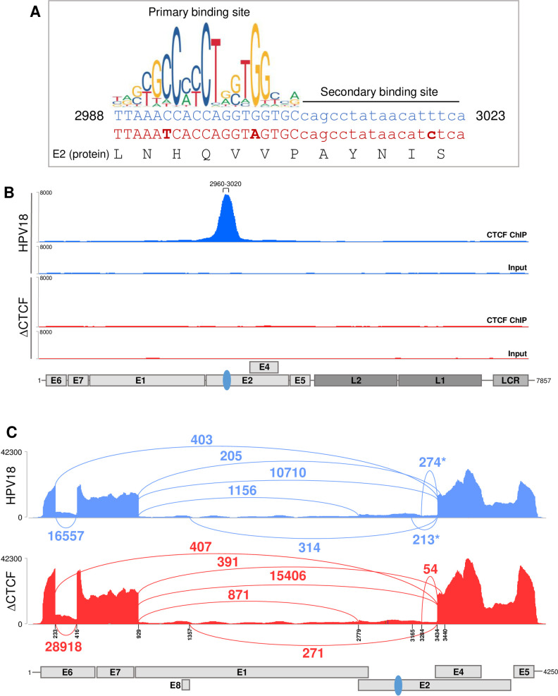
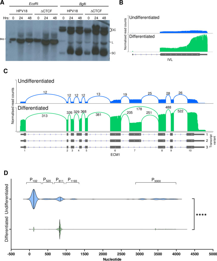
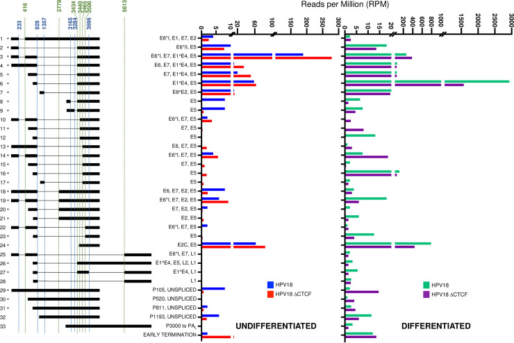
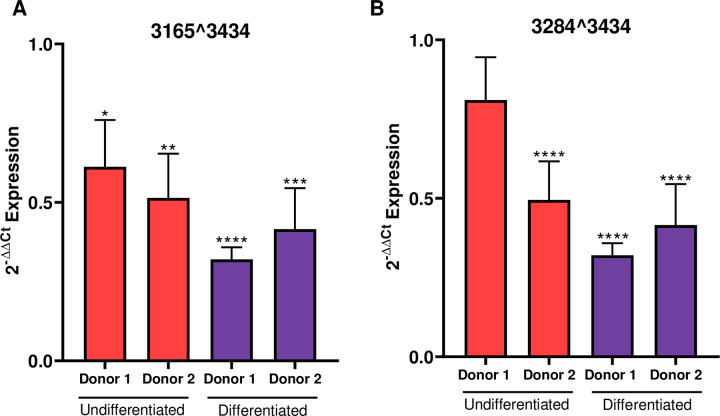
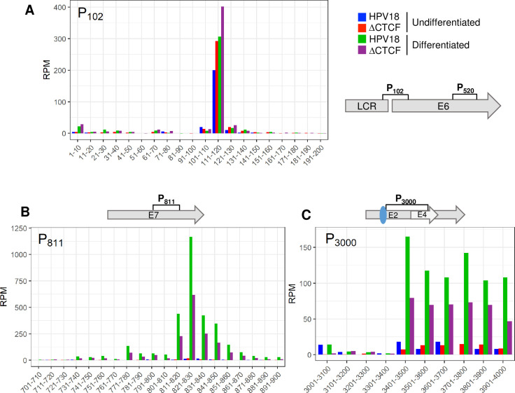
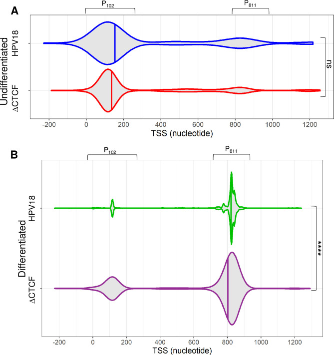
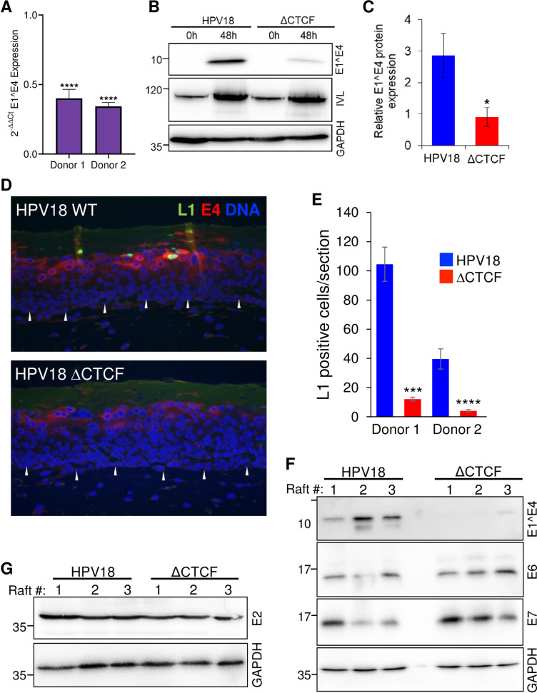
The ubiquitous host protein, CCCTC-binding factor (CTCF), is an essential regulator of cellular transcription and functions to maintain epigenetic boundaries, stabilise chromatin loops and regulate splicing of alternative exons. We have previously demonstrated that CTCF binds to the E2 open reading frame (ORF) of human papillomavirus (HPV) 18 and functions to repress viral oncogene expression in undifferentiated keratinocytes by co-ordinating an epigenetically repressed chromatin loop within HPV episomes. Keratinocyte differentiation disrupts CTCF-dependent chromatin looping of HPV18 episomes promoting induction of enhanced viral oncogene expression. To further characterise CTCF function in HPV transcription control we utilised direct, long-read Nanopore RNA-sequencing which provides information on the structure and abundance of full-length transcripts. Nanopore analysis of primary human keratinocytes containing HPV18 episomes before and after synchronous differentiation allowed quantification of viral transcript species, including the identification of low abundance novel transcripts. Comparison of transcripts produced in wild type HPV18 genome-containing cells to those identified in CTCF-binding deficient genome-containing cells identifies CTCF as a key regulator of differentiation-dependent late promoter activation, required for efficient E1^E4 and L1 protein expression. Furthermore, our data show that CTCF binding at the E2 ORF promotes usage of the downstream weak splice donor (SD) sites SD3165 and SD3284, to the dominant E4 splice acceptor site at nucleotide 3434. These findings demonstrate that in the HPV life cycle both early and late virus transcription programmes are facilitated by recruitment of CTCF to the E2 ORF.


Conflict of interest statement

The authors have declared that no competing interests exist.

Figures

Fig 1
Fig 1. Abrogation of CTCF recruitment to the HPV18 E2 ORF alters early transcript splicing.
(A) Nucleotide sequence of the CTCF binding site identified in the E2 ORF (nucleotides 2988–3023; blue text). The primary and secondary CTCF binding sites are shown as detailed in [46]. Conservative nucleotide substitutions introduced in ΔCTCF-HPV18 mutant (red text) are shown in bold. The E2 protein sequence (black text) is unaltered. Graphical representation of the primary CTCF binding site motif was obtained from JASPER2018 (http://jaspar.genereg.net/). (B) Enrichment of CTCF in the HPV18 genome was assessed by ChIP-Seq in either HPV18 (blue) or ΔCTCF-HPV18 (red) genome-containing keratinocytes. Next generation sequencing data were visualised using IGV. The position of HPV18 ORFs, LCR and E2-CTCF binding site (blue oval) are indicated below the alignment profiles. (C) Exon-exon junctions in Illumina RNA-Seq data sets of either HPV18 (blue) or ΔCTCF-HPV18 (red) genome-containing keratinocytes were identified and quantified in IGV and represented in Sashimi plots. The co-ordinates of splice donor and acceptor sites and annotated ORFs are indicated. The number of reads at each exon-exon junction is indicated. *denotes splicing event identified in HPV18 but reduced or not detected in ΔCTCF-HPV18 genome containing cells.
Fig 2
Fig 2. Analysis of differentiation-dependent host cell gene expression and HPV transcriptional start site usage.
HPV18-HFK were synchronously differentiated in methylcellulose for 48 hrs. (A) Amplification of HPV18 and ΔCTCF-HPV18 episomes was detected by Southern blotting following digestion with EcoRI to linearise the HPV18 episomes, or BglII which digests cellular DNA only (OC, open circle; L, linear; SC, supercoiled). (B-D) Host and viral transcriptomes in undifferentiated (blue) and differentiated (green) HPV18-HFK were analysed by long read Nanopore RNA-Seq, demonstrating enhanced involucrin (IVL) expression following keratinocyte differentiation (B) and enhanced ECM1 expression combined with differentiation-induced exon 7 skipping in transcript variant 3; exon numbering and transcript variants are indicated to the right and below the ECM1 gene annotation. (D) Clustered HPV18 promoter usage in undifferentiated and differentiated keratinocytes showing differentiation-dependent alteration of the major early (P102) and major late (P811) promoter usage. ****p < 0.0001 (Fisher’s test).
Fig 3
Fig 3. Quantitative analysis of the HPV18 transcriptome in undifferentiated and differentiated keratinocytes and alterations induced by abrogation of CTCF binding.
Alignment of Nanopore direct RNA sequencing data to the HPV18 genome facilitated the characterisation of all HPV-specific transcripts. Transcripts were included in the data set if they were represented by three or more individual reads in at least two samples. The relative abundance of each transcript type was calculated in reads per million (RPM) of the total reads in each sample. Relative abundance (RPM) of each transcript is shown for HPV18 (blue) and ΔCTCF-HPV18 (red) genome-containing cells in undifferentiated keratinocytes (left) and for HPV18 (green) and ΔCTCF-HPV18 (purple) in differentiated keratinocytes (right). Splice donor (blue) and acceptor (green) sites are indicated above the transcript map and HPV18 ORFs encoded by each transcript are shown. *denotes transcripts that have previously been identified [18, 19].
Fig 4
Fig 4. Loss of CTCF binding at the E2-ORF of HPV18 causes reduced downstream transcript splicing.
Splicing at (A) 3165^3434 and (B) 3284^3434 was assessed by qRT-PCR in two independent keratinocyte donors cultured in monolayer (undifferentiated; red) and in methylcellulose for 48 hrs (differentiated; purple). Data shown are the mean and standard error of transcript abundance normalised to β-actin and expressed and fold expression (2-ΔΔCT) compared to donor matched HPV18 episome containing cells.
Fig 5
Fig 5. Quantitative analysis of transcription start site usage in undifferentiated and differentiated keratinocytes and CTCF-dependent regulation of promoter activity.
The 5’ end of each HPV18 transcript was identified in Nanopore RNA sequencing data sets and relative abundance calculated as reads per million (RPM). Total counts at each nucleotide position were binned into 10 (A and B) or 100 (D) nucleotide regions in the data shown. Transcripts originating around the P102 (A), P811 (B) and P3000 (C) promoters were identified in HPV18 and ΔCTCF-HPV18 cells in undifferentiated (blue and red, respectively) and methylcellulose differentiated (green and purple, respectively) cultures. Relevant HPV18 genome features are shown alongside each panel. The E2-CTCF binding site is indicated by a blue oval.
Fig 6
Fig 6. CTCF regulates efficient differentiation-dependent HPV18 late promoter activation.
The 5’ end of each viral transcript was identified and the distribution shown in violin plots in (A) undifferentiated and (B) differentiated keratinocytes containing HPV18 (blue and green, respectively) and ΔCTCF-HPV18 (red and purple, respectively) episomes. Data distribution are shown by the kernel shape and median indicated with a vertical solid line. The widest sections of each violin plot indicates the highest probability of promoter usage within that region of the HPV18 genome. The shape of the distribution indicates the concentration of data points in a particular region; the steeper the side of each bubble indicates a greater concentration of data points. ns, non significant; ****p<0.0001 (Fisher’s test).
Fig 7
Fig 7. Abrogation of CTCF binding to the HPV18 genome causes a significant reduction in differentiation-dependent late protein abundance.
(A) HPV18 genome containing keratinocytes (HPV18 or ΔCTCF-HPV18) grown in monolayer (undifferentiated, 0h) or differentiated in methylcellulose (48h) and E1^E4, involucrin (IVL) and GAPDH protein expression analysed by Western blotting. (B) Relative E1^E4 protein expression in comparison to GAPDH was quantified in three independent experiments by densitometry. Data are the mean +/- standard deviation. * denotes p<0.05. (C) E1^E4 (red) and L1 (green) protein abundance was analysed by indirect immunofluorescence in epithelia derived from HPV18 and ΔCTCF-HPV18 genome-containing keratinocytes grown in organotypic raft culture. Cellular nuclei are shown in blue, and the basal layer indicated with white arrows. Scale bar indicates 10 μm. (D) The total number of L1 positive cells per section of three independent raft cultures grown from two independent keratinocyte donors was counted. Data show the mean +/- standard deviation. *** p<0.001, **** p<0.0001. (E) E1^E4, E6 and E7, and (F) E2 protein expression in organotypic raft cultures was assessed by Western blotting lysates harvested from three independent raft cultures alongside GADPH loading control. Molecular weight markers are indicated on the left of Western blots (kDa).
Fig 8
Fig 8. Keratinocyte differentiation induces increased H4Ac abundance at the HPV18 late promoter in wild type but not ΔCTCF-HPV18 genome-containing cells.
HPV18 and HPV18-ΔCTCF genome-containing primary keratinocytes grown in monolayer (undifferentiated; blue and green, respectively) or differentiated in methylcellulose-containing media for 48 hrs (green and purple, respectively). Enrichment of H4Ac was assessed by ChIP-qPCR. Each bar in the chart represents the mid-point for primer pairs used to amplify immunoprecipitated chromatin. Fold binding over IgG control was calculated. The data shown are the mean and standard deviation of three independent replicates. Annotation of the HPV18 LCR, promoters, ORFs and CTCF binding site (blue oval) is provided below.

References

    1. Tommasino M. The human papillomavirus family and its role in carcinogenesis. Semin Cancer Biol. 2014;26:13–21. doi: 10.1016/j.semcancer.2013.11.002 . - DOI - PubMed
    1. Stunkel W, Bernard HU. The chromatin structure of the long control region of human papillomavirus type 16 represses viral oncoprotein expression. J Virol. 1999;73(3):1918–30. doi: 10.1128/JVI.73.3.1918-1930.1999 ; PubMed Central PMCID: PMC104433. - DOI - PMC - PubMed
    1. Hummel M, Lim HB, Laimins LA. Human papillomavirus type 31b late gene expression is regulated through protein kinase C-mediated changes in RNA processing. J Virol. 1995;69(6):3381–8. doi: 10.1128/JVI.69.6.3381-3388.1995 ; PubMed Central PMCID: PMC189050. - DOI - PMC - PubMed
    1. Ruesch MN, Stubenrauch F, Laimins LA. Activation of papillomavirus late gene transcription and genome amplification upon differentiation in semisolid medium is coincident with expression of involucrin and transglutaminase but not keratin-10. J Virol. 1998;72(6):5016–24. doi: 10.1128/JVI.72.6.5016-5024.1998 ; PubMed Central PMCID: PMC110064. - DOI - PMC - PubMed
    1. Wooldridge TR, Laimins LA. Regulation of human papillomavirus type 31 gene expression during the differentiation-dependent life cycle through histone modifications and transcription factor binding. Virology. 2008;374(2):371–80. doi: 10.1016/j.virol.2007.12.011 ; PubMed Central PMCID: PMC2410142. - DOI - PMC - PubMed

Publication types