

[iOCT in clinical use : Correlation of intraoperative morphology and postoperative visual outcome in patients with full thickness macular hole]
- PMID: 34735612
- PMCID: PMC9076724
- DOI: 10.1007/s00347-021-01527-w
[iOCT in clinical use : Correlation of intraoperative morphology and postoperative visual outcome in patients with full thickness macular hole]
Abstract
Background: Due to intraoperative optical coherence tomography (iOCT), observation of retinal morphological changes during surgery has become possible.
Objective: To analyze the intraoperative morphology of full thickness macular holes (FTMH) and the correlation with the postoperative function, a retrospective, observational clinical study was performed analyzing 32 eyes of patients treated at the hospital of the technical university of Munich.
Material and methods: Using iOCT in 32 eyes of 32 consecutive patients, the operative morphology was analyzed during surgery. These findings were then correlated with the postoperative visual outcome.
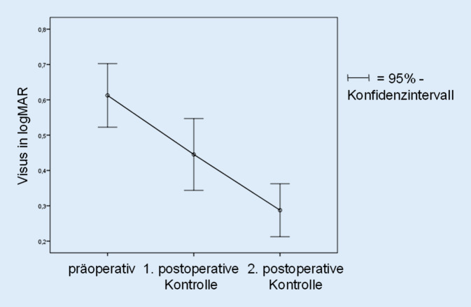
Results: After posterior vitreous detachment (PVD) the macular hole index (MHI) decreased by -0.05 (p = 0.01) and the base diameter (BD) increased by +99.4 μm (SD = 197.8 μm; p = 0.04). Closure rate was 100% at the first visit after a mean time of 73 days and the postoperative best corrected visual acuity (BCVA) significantly improved (p < 0.05). There were significant correlations between intraoperative morphology and postoperative results indicating a relation between low MHI and better postoperative BCVA (SCC = 0.50; p = 0.02), large BD and better postoperative BCVA (SCC = 0.43; p = 0.05) and large aperture after PVD and higher improvement of BCVA (SCC = 0.44; p = 0.03).
Conclusion: Flattening and broadening of the FTMH occurred as a result of reduction of vitreoretinal traction. The significant correlation between a large operative BD and improved BCVA reveals the importance of intraoperative retinal relaxation.
Zusammenfassung: HINTERGRUND: Die Beurteilung der intraoperativen Veränderung der Netzhautmorphologie, insbesondere des vitreoretinalen Überganges, ist mithilfe der intraoperativen optischen Kohärenztomografie (iOCT) möglich geworden.
Ziel der arbeit: Um die Bedeutung der intraoperativen Morphologie beim durchgreifenden Makulaforamen (MF) für das postoperative funktionelle Ergebnis zu evaluieren, wurde eine retrospektive, klinische Beobachtungsstudie durchgeführt.
Material und methoden: Die Netzhautmorphologie wurde in 32 Augen von 32 konsekutiven Patienten mit durchgreifendem Makulaforamen mittels iOCT zu verschiedenen Zeitpunkten während der Operation beobachtet. Die Veränderungen wurden anschließend mit dem postoperativen funktionellen Ergebnis korreliert.
Ergebnisse: Nach Induktion der hinteren Glaskörperabhebung (HGA) reduzierte sich der Makulaforamen-Index (MHI) um −0,05 (p = 0,01), die basale Foramenbreite (FB) stieg um +99,4 μm (SD = 197,8 μm; p = 0,04). Die Verschlussrate betrug 100 % zum Zeitpunkt der ersten postoperativen Vorstellung nach im Mittel 73 Tagen, der postoperative Visus verbesserte sich signifikant (p < 0,05). Es zeigte sich eine signifikant positive Korrelation von intraoperativer Morphologie und postoperativem Ergebnis zwischen einem niedrigen MHI und einem besseren postoperativen Visus (SKK = 0,50; p = 0,02), zwischen einer großen FB und einem besseren postoperativen Visus (SKK = 0,43; p = 0,05) sowie zwischen einer breiten Apertur nach HGA und einem größeren Visusanstieg postoperativ (SKK = 0,44; p = 0,03).
Diskussion: Wir konnten eine Abflachung sowie eine Verbreiterung des MF durch Lösen der vitreoretinalen Zugkräfte beobachten. Aufgrund des Zusammenhangs zwischen einer großen intraoperativen FB mit einem besseren postoperativen Visus scheint die intraoperative Relaxierung der Netzhaut bedeutsam.
Keywords: IOCT; Macular Hole Surgery; Operative Imaging; SD-OCT; Vitreoretinal Surgery.
© 2021. The Author(s).


Similar articles
-
[Vitrectomy and iOCT-assisted inverted ILM flap technique in patients with full thickness macular holes].Ophthalmologe. 2019 Jul;116(7):617-624. doi: 10.1007/s00347-018-0769-y. Ophthalmologe. 2019. PMID: 30105564 German.
-
Surgically Induced Macular Detachment for Treatment of Refractory Full-Thickness Macular Hole: Anatomical and Functional Results.Ophthalmologica. 2019;242(2):98-105. doi: 10.1159/000500573. Epub 2019 Jun 20. Ophthalmologica. 2019. PMID: 31220838
-
Prediction of postoperative visual outcome based on hole configuration by optical coherence tomography in eyes with idiopathic macular holes.Am J Ophthalmol. 2004 Nov;138(5):709-16. doi: 10.1016/j.ajo.2004.04.063. Am J Ophthalmol. 2004. PMID: 15531303
-
[Intraoperative optical coherence tomography in vitreoretinal surgery].Vestn Oftalmol. 2023;139(5):113-120. doi: 10.17116/oftalma2023139051113. Vestn Oftalmol. 2023. PMID: 37942605 Review. Russian.
-
Intraoperative Optical Coherence Tomography in the Management of Macular Holes: State of the Art and Future Perspectives.Biomedicines. 2022 Nov 9;10(11):2873. doi: 10.3390/biomedicines10112873. Biomedicines. 2022. PMID: 36359392 Free PMC article. Review.
Cited by
-
[10 years of screening for retinopathy of prematurity (2009-2019) : Results analysis of two German level-1 neonatal intensive care units (NICUs) with university on-site screening and a telemedical approach in the non-university NICU].Ophthalmologie. 2023 Sep;120(9):920-931. doi: 10.1007/s00347-023-01847-z. Epub 2023 Apr 21. Ophthalmologie. 2023. PMID: 37083751 German.
-
Microstructural morphology and visual acuity outcome in eyes with epiretinal membrane before, during, and after membrane peeling in intraoperative optical coherence tomography assisted macular surgery.Int J Ophthalmol. 2023 May 18;16(5):748-754. doi: 10.18240/ijo.2023.05.12. eCollection 2023. Int J Ophthalmol. 2023. PMID: 37206168 Free PMC article.
References
-
- Bleidißel N, Friedrich J, Klaas J, et al. Inverted internal limiting membrane flap technique in eyes with large idiopathic full-thickness macular hole: long-term functional and morphological outcomes. Graefes Arch Clin Exp Ophthalmol. 2021;259(7):1759–1771. doi: 10.1007/s00417-021-05082-7. - DOI - PMC - PubMed
-
- Carl Zeiss Meditec Technische Daten (2017) Available from: https://www.zeiss.de/meditec/produkte/ophthalmologie/katarakt/visualisie.... Zugegriffen: 13.11.2017
Publication types
MeSH terms
LinkOut - more resources
Full Text Sources
Research Materials
