Comparative Study
. 2021 Nov 9;118(45):e2110996118.
doi: 10.1073/pnas.2110996118.

In-cell structures of conserved supramolecular protein arrays at the mitochondria-cytoskeleton interface in mammalian sperm

Affiliations
Comparative Study

In-cell structures of conserved supramolecular protein arrays at the mitochondria-cytoskeleton interface in mammalian sperm

Miguel Ricardo Leung et al. Proc Natl Acad Sci U S A. .

Abstract

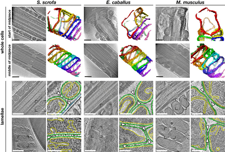
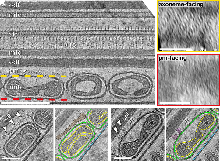
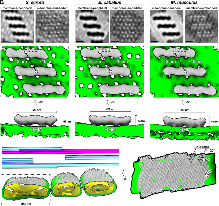
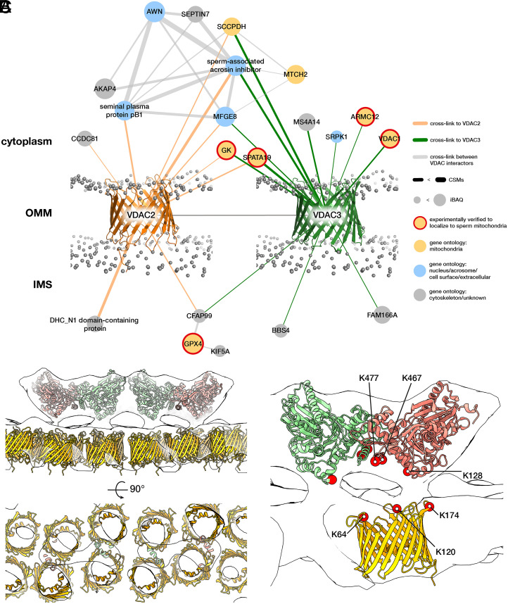
Mitochondria-cytoskeleton interactions modulate cellular physiology by regulating mitochondrial transport, positioning, and immobilization. However, there is very little structural information defining mitochondria-cytoskeleton interfaces in any cell type. Here, we use cryofocused ion beam milling-enabled cryoelectron tomography to image mammalian sperm, where mitochondria wrap around the flagellar cytoskeleton. We find that mitochondria are tethered to their neighbors through intermitochondrial linkers and are anchored to the cytoskeleton through ordered arrays on the outer mitochondrial membrane. We use subtomogram averaging to resolve in-cell structures of these arrays from three mammalian species, revealing they are conserved across species despite variations in mitochondrial dimensions and cristae organization. We find that the arrays consist of boat-shaped particles anchored on a network of membrane pores whose arrangement and dimensions are consistent with voltage-dependent anion channels. Proteomics and in-cell cross-linking mass spectrometry suggest that the conserved arrays are composed of glycerol kinase-like proteins. Ordered supramolecular assemblies may serve to stabilize similar contact sites in other cell types in which mitochondria need to be immobilized in specific subcellular environments, such as in muscles and neurons.

Keywords: cross-linking mass spectrometry; cryo-FIB milling; cryoelectron tomography; mitochondria–cytoskeleton contacts; subtomogram averaging.


Conflict of interest statement

The authors declare no competing interest.

Figures

Fig. 1.
Fig. 1.
Mitochondrial dimensions and cristae organization vary across species. (AF) Slices through VPP cryotomograms (Left) and corresponding three-dimensional segmentations (Right) of mitochondria from the start (AC) or middle (DF) of the midpiece from pig (A and D), horse (B and E), and mouse (C and F) sperm. (GL) Slices through cryotomograms of FIB-milled pig (G and H), horse (I and J), and mouse (K and L) sperm midpieces. Right panels show digital zooms of the regions boxed out in the Left panels. The OMM is traced in green, the inner mitochondrial membrane in yellow, and the plasma membrane in blue. Arrowheads indicate intermitochondrial linker complexes. Labels: nuc, nucleus; sc, segmented columns; m, mitochondria; odf, outer dense fibers; dc, distal centriole; ax, axoneme; mtd, microtubule doublets; cpa, central pair apparatus; and pm, plasma membrane. (Scale bars: [AL] Left panels, 250 nm, and [GL] Right panels, 100 nm.)
Fig. 2.
Fig. 2.
Ordered protein arrays on the OMM interact with the cytoskeleton. (A) Slice through a cryotomogram of an FIB-milled, horse sperm midpiece showing mitochondria (mito), the submitochondrial reticulum (smr) ODFs (odf), microtubule doublets (mtd), and the central pair apparatus (cpa). Note how individual complexes (like the radial spoke, rs) are visible in the raw tomogram. The ordered protein array is only found on the axoneme-facing surface (yellow) of midpiece mitochondria and not on the plasma membrane–facing surface (red). (B and C) Slices through a cryotomogram of an FIB-milled, horse sperm midpiece showing how the array directly interacts with the submitochondrial reticulum to anchor mitochondria to the flagellar cytoskeleton (arrowheads). In Right panels, the OMM is traced in green, the inner mitochondrial membrane in yellow, and the plasma membrane in blue. (Scale bars: [A] Left, 250 nm, Insets, 100 nm, and [B and C] 100 nm.)
Fig. 3.
Fig. 3.
Ordered protein arrays at the mitochondria–cytoskeleton interface are conserved across species. (AC) Subtomogram averages of the protein arrays and underlying OMM after applying twofold symmetry (note that density is black). (DF) Isosurface renderings of the subtomogram averages in AC with boat-shaped particles in gray and the OMM in green. (G, Left) Segmentation of the tomogram shown in Fig. 2A, with the OMM in green, the IMM in yellow, microtubule doublets in blue, and the cpa in pink. Subtomogram averages of boat-shaped particles are colored gray and plotted back into their positions and orientations in the tomogram. (Right) Rotated and zoomed-in view of the axoneme-facing surface of a mitochondrion. The axoneme is oriented horizontally, so the ladder-like arrays are oriented ∼120° to the flagellar long axis, and individual particles within the array are oriented ∼60° to this axis. (Scale bars, 10 nm.)
Fig. 4.
Fig. 4.
Modeling the OMM array as GK proteins anchored on VDACs. (A) The VDAC2/VDAC3 interactome derived from in-cell XL-MS of pig sperm. Protein nodes are colored according to their known subcellular localizations (yellow with red border, experimentally verified to localize to sperm mitochondria; yellow, gene ontology: mitochondria; blue, gene ontology: nucleus/acrosome/cell surface; and gray, gene ontology: cytoskeleton/unknown). Gray spheres indicate the phosphate groups of a simulated lipid bilayer which was structurally aligned based on the simulation for monomeric mouse VDAC1 (PDB 4C69) obtained from the MemProtMD server (97). (B) Modeling the OMM array as GK-like proteins anchored on VDACs. A GK-like dimer-of-dimers homology model (red and green) and VDAC2 homology models (yellow) were fitted into the pig subtomogram average map (white). (C) The positions of cross-linked Lys residues (red circles) are consistent with GK and VDAC orientation assignments in our model. Note that the cross-links are shown for VDAC2, but we detect an additional cross-link between K140 on GK and VDAC3 (SI Appendix, Fig. S6).

Comment in

References

    1. Viana M. P., et al. ., Mitochondrial fission and fusion dynamics generate efficient, robust, and evenly distributed network topologies in budding yeast cells. Cell Syst. 10, 287–297.e5 (2020). - PubMed
    1. Glancy B., et al. ., Mitochondrial reticulum for cellular energy distribution in muscle. Nature 523, 617–620 (2015). - PMC - PubMed
    1. Vincent A. E., Turnbull D. M., Eisner V., Hajnóczky G., Picard M., Mitochondrial nanotunnels. Trends Cell Biol. 27, 787–799 (2017). - PMC - PubMed
    1. Moore A. S., Holzbaur E. L. F., Mitochondrial-cytoskeletal interactions: Dynamic associations that facilitate network function and remodeling. Curr. Opin. Physiol. 3, 94–100 (2018). - PMC - PubMed
    1. Fenton A. R., Jongens T. A., Holzbaur E. L. F., Mitochondrial dynamics: Shaping and remodeling an organelle network. Curr. Opin. Cell Biol. 68, 28–36 (2021). - PMC - PubMed

Publication types