. 2021 Nov 4;11(1):21328.
doi: 10.1038/s41598-021-00724-6.

Desiccation-induced fibrous condensation of CAHS protein from an anhydrobiotic tardigrade

Affiliations

Desiccation-induced fibrous condensation of CAHS protein from an anhydrobiotic tardigrade

Maho Yagi-Utsumi et al. Sci Rep. .

Abstract

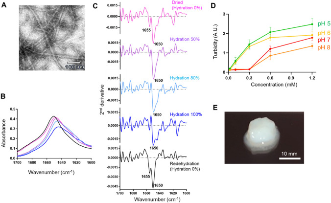
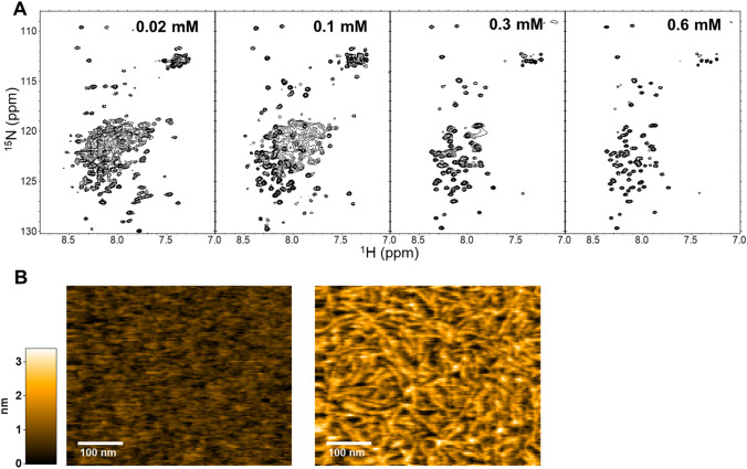
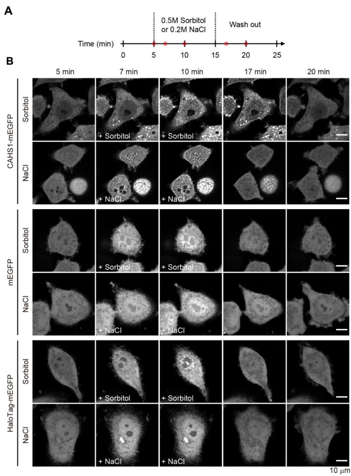
Anhydrobiosis, one of the most extensively studied forms of cryptobiosis, is induced in certain organisms as a response to desiccation. Anhydrobiotic species has been hypothesized to produce substances that can protect their biological components and/or cell membranes without water. In extremotolerant tardigrades, highly hydrophilic and heat-soluble protein families, cytosolic abundant heat-soluble (CAHS) proteins, have been identified, which are postulated to be integral parts of the tardigrades' response to desiccation. In this study, to elucidate these protein functions, we performed in vitro and in vivo characterizations of the reversible self-assembling property of CAHS1 protein, a major isoform of CAHS proteins from Ramazzottius varieornatus, using a series of spectroscopic and microscopic techniques. We found that CAHS1 proteins homo-oligomerized via the C-terminal α-helical region and formed a hydrogel as their concentration increased. We also demonstrated that the overexpressed CAHS1 proteins formed condensates under desiccation-mimicking conditions. These data strongly suggested that, upon drying, the CAHS1 proteins form oligomers and eventually underwent sol-gel transition in tardigrade cytosols. Thus, it is proposed that the CAHS1 proteins form the cytosolic fibrous condensates, which presumably have variable mechanisms for the desiccation tolerance of tardigrades. These findings provide insights into molecular strategies of organisms to adapt to extreme environments.


Conflict of interest statement

The authors declare no competing interests.

Figures

Figure 1
Figure 1
In vitro characterization of the fibrous condensation of CAHS1 proteins. (A) TEM image of CAHS1 protein fibrils under dry conditions. (B) Amide I band of the CAHS1 protein and (C) its second derivative under 0 (magenta), 50 (purple), 80 (cyan), and 100% hydration (D2O) and dehydration conditions. After 100% hydration (blue), the sample was measured again under redehydration condition (black). (D) Turbidity (absorbance at 595 nm) of the CAHS1 protein at 25 °C and pH 5 (green), pH 6 (yellow), pH 7 (red), and pH 8 (orange). The error bars show the standard deviation of three replicates. (E) The CAHS1 protein hydrogel formed in 20 mM potassium phosphate buffer (pH 6.0) at 25 °C. The initial protein concentration was 1.2 mM.
Figure 2
Figure 2
Fibril formation of the CAHS1 protein in vitro. (A) 1H-15N HSQC spectra of the CAHS1 protein at protein concentrations of 0.02, 0.1, 0.3, and 0.6 mM. (B) Typical HS-AFM images of CAHS1 protein fibrils. Left: the HS-AFM image of the mica surface with CAHS1 protein at a final concentration of 0.4 μM, about 3 min after injection into the observation solution. Right: the HS-AFM image about 2 min after the addition of CAHS1 protein at a final concentration of 3.3 μM.
Figure 3
Figure 3
Real-time monitoring of the reversible formation of CAHS1 protein particles. (A) Timeline of time-laps imaging with hyperosmotic shock. The red dots represent the time points when the representative cells are presented in panel (B). (B) Time-laps images of HeLa cells overexpressing the CAHS1-mEGFP, mEGFP, and Halo Tag-mEGFP proteins. A sorbitol or sodium chloride solution was added at 5 min. After 10 min, the cells were washed with a flesh medium using a microfluidics system.

References

    1. Hibshman JD, Clegg JS, Goldstein B. Mechanisms of desiccation tolerance: Themes and variations in brine shrimp, roundworms, and tardigrades. Front. Physiol. 2020;11:592016. doi: 10.3389/fphys.2020.592016. - DOI - PMC - PubMed
    1. Rapoport A, Golovina EA, Gervais P, Dupont S, Beney L. Anhydrobiosis: Inside yeast cells. Biotechnol. Adv. 2019;37:51–67. doi: 10.1016/j.biotechadv.2018.11.003. - DOI - PubMed
    1. Janis B, Belott C, Menze MA. Role of intrinsic disorder in animal desiccation tolerance. Proteomics. 2018;18:e1800067. doi: 10.1002/pmic.201800067. - DOI - PubMed
    1. Garcia AH. Anhydrobiosis in bacteria: From physiology to applications. J. Biosci. 2011;36:939–950. doi: 10.1007/s12038-011-9107-0. - DOI - PubMed
    1. Guidetti R, Jönsson KI. Long-term anhydrobiotic survival in semi-terrestrial micrometazoans. J. Zool. Lond. 2002;257:181–187. doi: 10.1017/S095283690200078X. - DOI

Publication types