. 2021 Nov 4;11(1):21642.
doi: 10.1038/s41598-021-01004-z.

Claudin-9 constitutes tight junctions of folliculo-stellate cells in the anterior pituitary gland

Affiliations

Claudin-9 constitutes tight junctions of folliculo-stellate cells in the anterior pituitary gland

Atsuko Y Higashi et al. Sci Rep. .

Abstract

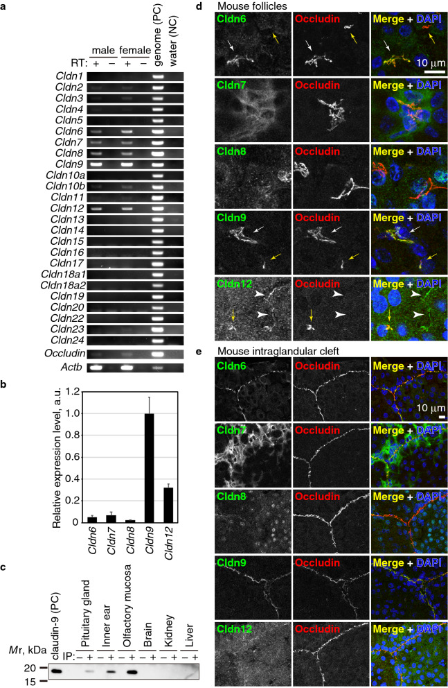
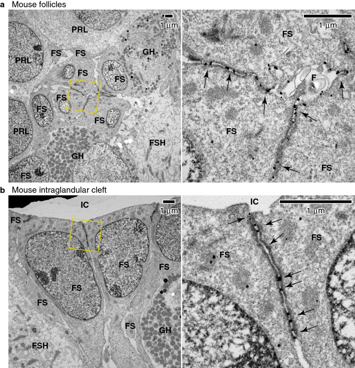
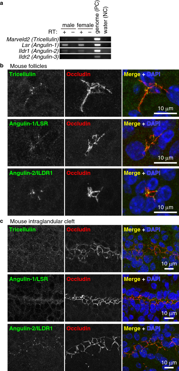
The anterior pituitary gland regulates growth, metabolism, and reproduction by secreting hormones. Folliculo-stellate (FS) cells are non-endocrine cells located among hormone-producing cells in the anterior pituitary glands. They form follicular lumens, which are sealed by tight junctions (TJs). Although FS cells are hypothesized to contribute to fine-tuning of endocrine cells, little is known about the exact roles of FS cells. Here, we investigated the molecular composition of TJs in FS cells. We demonstrated that occludin is a good marker for TJs in the pituitary gland and examined the structure of the lumens surrounded by FS cells. We also found that claudin-9 is a major component of TJs in the FS cells. In immunoelectron microscopy, claudin-9 was specifically localized at TJs of the FS cells. The expression of claudin-9 was gradually increased in the pituitary gland after birth, suggesting that claudin-9 is developmentally regulated and performs some specific functions on the paracellular barrier of follicles in the pituitary gland. Furthermore, we found that angulin-1, angulin-2, and tricellulin are localized at the tricellular contacts of the FS cells. Our findings provide a first comprehensive molecular profile of TJs in the FS cells, and may lead us towards unveiling the FS cell functions.


Conflict of interest statement

The authors declare no competing interests.

Figures

Figure 1
Figure 1
Anterior pituitary gland and FS cells. (a) Schematic illustration of the mouse pituitary gland. (b) Whole-mount staining of the pituitary gland of a mouse neonate (P2) using anti-occludin mAb. (c), (d) Immunostaining of frozen sections of the adult mouse (c) or adult rat (d) anterior pituitary gland using anti-occludin mAb, anti-S100 pAb, and DAPI.
Figure 2
Figure 2
Occludin-positive cells are FS cells. (a) Triple-staining of the adult mouse anterior pituitary gland using anti-occludin mAb, anti-VE-cadherin pAb and anti-ZO-1 pAb. Note that occludin stains the ZO-1-positive FS cells (white arrows) and does not stain the endothelial cells labeled with both VE-cadherin and ZO-1 (yellow arrows). (b), (c) Immunostaining of the adult mouse (b) or rat (c) anterior pituitary gland using anti-GH, LH, TSH, Prolactin or ACTH pAbs and anti-occludin mAb. Note that occludin does not colocalize with any hormone signals.
Figure 3
Figure 3
Claudin-9 is a component of FS cells. (a) RT-PCR of the pituitary gland of adult male and female mice. Since the forward and reverse primers were designed in the same exon, the genome DNA was used as a positive control (PC) and water was used as a negative control (NC). The reaction mix without reverse transcriptase (RT) also served as a negative control. (b) Real-time RT-PCR of the adult mouse pituitary gland. The expression levels were normalized using known concentration of DNA fragments as standards. The expression level of cldn9 was set to 1. Error bars indicate s.d. (c) Immunoprecipitation of claudin-9 from adult mouse organs using rat anti-claudin-9 (clone 1B1) and immunoblotting using goat anti-claudin-9 pAb. Human CLDN9-expressing HEK293T cell lysate served as a positive control (PC). Note that the claudin-9 band was detected in the pituitary gland lysate as well as in the inner ear and olfactory mucosa. (d), (e) Immunostaining of the adult mouse anterior pituitary gland using anti-claudin-6 pAb, anti-claudin-7 pAb, anti-claudin-8 pAb, anti-claudin-9 mAb (clone 1B1), or anti-claudin-12 pAb together with anti-occludin mAb or pAb and DAPI.
Figure 4
Figure 4
Claudin-9 is localized at the TJs of FS cells. (a,b) Immunoelectron microscopy of the adult mouse anterior pituitary gland. Claudin-9 signal was detected at TJs (arrows) between the FS cells (FS) in the follicles (F) (a) and the intraglandular cleft (IC) (b). FSH, FSH cells; PRL, PRL cells; GH, GH cells.
Figure 5
Figure 5
Tricellulin, angulin-1, and angulin-2 are expressed in FS cells. (a) RT-PCR of the pituitary gland of adult male and female mice. (b), (c) Immunostaining of the adult mouse anterior pituitary gland using anti-tricellulin mAb (green) and anti-occludin pAb (red), anti-angulin-1 pAb (green) and anti-occludin mAb (red), and anti-angulin-2 pAb (green) and anti-occludin mAb (red) together with DAPI (blue).
Figure 6
Figure 6
Anti-claudin-9 is applicable for immunohistochemistry. (a) Immunocytochemical staining of FFPE samples of HEK293T cells expressing human CLDN9 using anti-claudin-9 mAbs (clones 1B1 and 1B8) and anti-claudin-9 pAb. (b) Immunohistochemical staining of FFPE samples of the human pituitary gland using anti-claudin-9 mAb (clone 1B1) and anti-claudin-9 pAb.

References

    1. Rinehart JF, Farquhar MG. Electron microscopic studies of the anterior pituitary gland. J. Histochem. Cytochem. Off. J. Histochem. Soc. 1953;1:93–113. doi: 10.1177/1.2.93. - DOI - PubMed
    1. Farquhar MG. Corticotrophs of the rat adenohypophysis as revealed by electron microscopy. Anat. Rec. 1957;127:291.
    1. Le Tissier PR, et al. Anterior pituitary cell networks. Front. Neuroendocrinol. 2012;33:252–266. doi: 10.1016/j.yfrne.2012.08.002. - DOI - PubMed
    1. Cocchia D, Miani N. Immunocytochemical localization of the brain-specific S-100 protein in the pituitary gland of adult rat. J. Neurocytol. 1980;9:771–782. doi: 10.1007/BF01205018. - DOI - PubMed
    1. Nakajima T, Yamaguchi H, Takahashi K. S100 protein in folliculostellate cells of the rat pituitary anterior lobe. Brain Res. 1980;191:523–531. doi: 10.1016/0006-8993(80)91300-1. - DOI - PubMed

Publication types