Review
. 2021 Nov;10(13):e12161.
doi: 10.1002/jev2.12161.

Microbiota-derived extracellular vesicles in interkingdom communication in the gut

Affiliations
Review

Microbiota-derived extracellular vesicles in interkingdom communication in the gut

Natalia Díaz-Garrido et al. J Extracell Vesicles. 2021 Nov.

Abstract

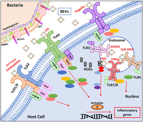
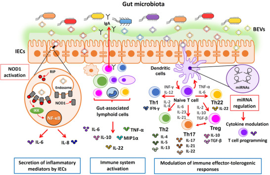
The intestine is fundamental in controlling human health. Intestinal epithelial and immune cells are continuously exposed to millions of microbes that greatly impact on intestinal epithelial barrier and immune function. This microbial community, known as gut microbiota, is now recognized as an important partner of the human being that actively contribute to essential functions of the intestine but also of distal organs. In the gut ecosystem, bidirectional microbiota-host communication does not involve direct cell contacts. Both microbiota and host-derived extracellular vesicles (EVs) are key players of such interkingdom crosstalk. There is now accumulating body of evidence that bacterial secreted vesicles mediate microbiota functions by transporting and delivering into host cells effector molecules that modulate host signalling pathways and cell processes. Consequently, vesicles released by the gut microbiota may have great influence on health and disease. Here we review current knowledge on microbiota EVs and specifically highlight their role in controlling host metabolism, intestinal barrier integrity and immune training.

Keywords: bacterial membrane vesicles; gut microbiota; gut permeability; immune regulation; intestinal homeostasis; probiotics.


Conflict of interest statement

AUTHOR CONTRIBUTIONS

Conceptualization, J.B and L.B; investigation, N.D.G, writing—original draft preparation, N.D.G.; writing—review and editing, L.B and J.B; supervision L.B; project administration, J.B; funding acquisition, L.B and J.B.

Figures

FIGURE 1
FIGURE 1
Recognition of BEV‐associated molecular patterns by host immune receptors. The drawing schematically shows toll‐like receptors (TLRs) located at the host cell membrane (TLR1/6, TLR2, TLR4, and TLR5), TLRs located at the endosomal membranes (TLR3, TLR7/8, TLR9) and cytosolic NODs (NOD1/NOD2), and the downstream signalling pathways that lead to activation of NF‐kB. Proteins of the different signalling pathways are drawn as coloured ellipses. The molecular associated pattern that specifically interacts with each receptor is indicated in red. LPS: lipopolysaccharide, PGN: peptidoglycan
FIGURE 2
FIGURE 2
Metabolic activities associated with Bacteroides‐derived BEVs known to contribute to gut ecology and food metabolism. (a) The enzymes included as cargo in B. thetaiotaomicron and B. fragilis BEVs responsible for metabolic effects are indicated. (b) The drawing shows the intestinal epithelial barrier and the underlying immune system. The mucin layer (in green) maintains segregation between luminal microbes and the intestinal epithelium. Blue boxes connect the vesicular enzyme (in red) with each specific function. SCFAs: short‐chain fatty acids, InsP6: inositol phosphates
FIGURE 3
FIGURE 3
Modulation of the gut epithelial barrier by microbiota BEVs. Schematic representation of the intestinal epithelium, where tight junction (TJ) proteins are indicated by coloured bars connecting adjacent intestinal epithelial cells (IECs). BEVs released by E. coli Nissle 1917 (EcN) and ECOR63 (left panel), or A. muciniphila (right panel) migrate through the inner mucus layer and reach the epithelium. Regulatory mechanisms influencing the integrity of the intestinal barrier known to be activated by BEVs are indicated below the drawing, specifying whether evidences were obtained from in vitro assays (culture of IECs) or in vivo models of increased intestinal permeability (experimental colitis and high fat diet (HFD)‐induced diabetic model). Upregulation/downregulation of gene transcription is indicated by red arrows. For each experimental model, the beneficial effects of BEVs counteracting disease alterations are shown by blue arrows. Overall, the regulatory effects mediated by microbiota BEVs result in gut epithelial barrier reinforcement and the subsequent reduction of intestinal permeability
FIGURE 4
FIGURE 4
Schematic picture summarizing the immunomodulatory effects elicited by microbiota‐derived BEVs in the gut. The drawing shows the intestinal epithelium covered with the mucin layer that prevents access of luminal microbes while allowing passage of BEVs. Immune cells (lymphocytes, macrophages and dendritic cells) in the lamina propria are shown below the epithelial monolayer. Microbiota derived BEVs exert immune modulation by two main mechanisms. (i) Undirect activation of immune cells through the intestinal epithelium (left scheme). Internalized EVs by intestinal epithelial cells (IECs) activate the cytosolic receptor NOD1 that triggers secretion of immune effectors, which in turn stimulate gut‐associated lymphoid cells to produce a wide range of cytokines. Activation of the NOD1 signalling pathway by microbiota BEVs is shown encircled in more detail. BEVs are internalized through clathrin‐mediated endocytosis and recruit NOD1 (grey cylinders) to early endosomes. Activated NOD1 interacts with the specific kinase RIP2 (red circles), which leads to NF‐kB activation and the subsequent upregulation of host genes involved in the inflammatory response (IL‐6, IL‐8). (ii) Direct activation of gut resident immune cells by microbiota BEVs that leads to secretion of immune mediators and secretory IgA (middle scheme). In addition to direct interaction with microbiota BEVs that reach the gut‐associated lymphoid tissue via transcytosis across M cells, dendritic cells (DCs) also interact with luminal BEVs by extending pseudopodia across the epithelial cell layer (right scheme). Studies with several gut microbiota species revealed that BEVs activate DCs in a strain‐specific manner. Differential regulation of miRNAs in DCs is one of the regulatory mechanisms involved in the specific immunomodulatory effects of BEVs isolated from probiotic and commensal E. coli is (highlighted in the adjacent violet circle). In conclusion, DCs integrate incoming signals delivered by microbiota BEVs and set up specific programs that promote differentiation of naïve T cells into effector T cells (Th1, Th2, Th17, Th22) or regulatory T cells (Treg), thus allowing coordination of suitable T cell responses
FIGURE 5
FIGURE 5
Mechanisms that mediate translocation of microbiota‐derived BEVs across the intestinal epithelium. The processes are numbered and indicated in red. BEVs released by gut microbes are taken up by intestinal epithelial cells (IECs) via endocytosis (1). Internalized BEVs can reach the basolateral membrane of IECs and be translocated into the lamina propria by transcytosis (2). In addition, the paracellular route (3) also allows passage of luminal BEVs to the underlying submucosa. This route is favoured under conditions of gut dysbiosis. Once in the lamina propria, translocated BEVs directly interact with resident gut immune cells, triggering suitable immune responses. Direct sampling and phagocytosis of luminal BEVs by DCs also mediate passage of luminal BEVs into the internal milieu (4). There is evidence that BEVs cross endothelial barriers and reach blood vessels, thus being distributed to distal organs

References

    1. Aguilera, L. , Toloza, L. , Gimenez, R. , Odena, A. , Oliveira, E. , Aguilar, J. , Badia, J. , & Baldoma, L. (2014). Proteomic analysis of outer membrane vesicles from the probiotic strain Escherichia coli Nissle 1917. Proteomics, 14(2–3), 222–229. 10.1002/pmic.201300328 - DOI - PubMed
    1. Ahmadi Badi, S. , Moshiri, A. , Ettehad Marvasti, F., Mojtahedzadeh, M. , Kazemi, V. , & Siadat, S. D. (2020). Extraction and evaluation of outer membrane vesicles from two important gut microbiota members, Bacteroides fragilis and Bacteroides thetaiotaomicron . Cell Journal, 22(3), 344–349. 10.22074/cellj.2020.6499 - DOI - PMC - PubMed
    1. Al‐Nedawi, K. , Mian, M. F. , Hossain, N. , Karimi, K. , Mao, Y.‐K. , Forsythe, P. , Min, K. K. , Stanisz, A. M. , Kunze, W. A. , & Bienenstock, J. (2015). Gut commensal microvesicles reproduce parent bacterial signals to host immune and enteric nervous systems. FASEB Journal : Official Publication of the Federation of American Societies for Experimental Biology, 29(2), 684–695. 10.1096/fj.14-259721 - DOI - PubMed
    1. Altindis, E. , Fu, Y. , & Mekalanos, J. J. (2014). Proteomic analysis of Vibrio cholerae outer membrane vesicles. Proceedings of the National Academy of Sciences of the United States of America, 111(15), E1548‐56. 10.1073/pnas.1403683111 - DOI - PMC - PubMed
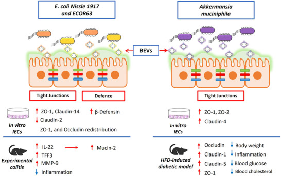
    1. Alvarez, C.‐S. , Giménez, R. , Cañas, M.‐A. , Vera, R. , Díaz‐Garrido, N. , Badia, J. , & Baldomà, L. (2019). Extracellular vesicles and soluble factors secreted by Escherichia coli Nissle 1917 and ECOR63 protect against enteropathogenic E. coli‐induced intestinal epithelial barrier dysfunction. BMC Microbiology, 19(1). 10.1186/s12866-019-1534-3 - DOI - PMC - PubMed

Publication types

MeSH terms

LinkOut - more resources