

Electrocardiogram evolution of acute anterior ST-segment elevation myocardial infarction following pericarditis
- PMID: 34738690
- PMCID: PMC8916549
- DOI: 10.1111/anec.12906
Electrocardiogram evolution of acute anterior ST-segment elevation myocardial infarction following pericarditis
Abstract
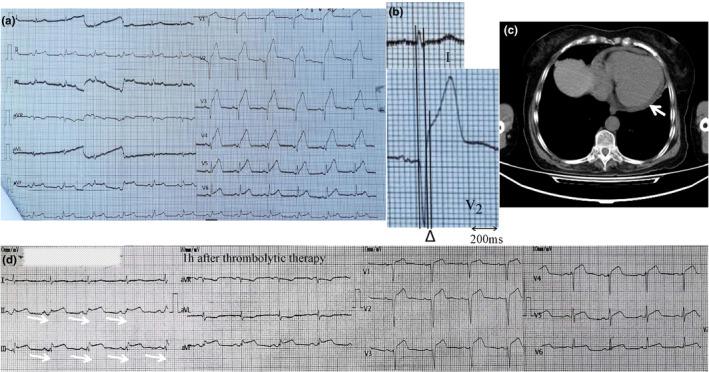
Electrocardiogram is a powerful tool for differentiating acute ST-segment elevation myocardial infarction (STEMI) and pericarditis. However, an unusual ECG presentation of the simultaneous occurrence of the two conditions has not been reported previously. In this article, we report a case of ECG evolution of acute anterior STEMI following pericarditis with pericardial effusion (PE) and find that QRS complex widening in ECG lead with maximal ST-segment elevation is also applicable for identifying STEMI even in patients with prior pericarditis. Undoubtedly, our case can help prevent emergency physicians from making incorrect diagnoses and administering inappropriate treatments.
Keywords: ECG evolution; ST-segment elevation myocardial infarction; pericarditis.
© 2021 The Authors. Annals of Noninvasive Electrocardiology published by Wiley Periodicals LLC.
Conflict of interest statement
There are no conflicts of interest that should be stated.
Figures


References
-
- Bozbeyoglu, E. , Yildirimturk, O. , Aslanger, E. , Simsek, B. , Karabay, C. Y. , Ozveren, O. , & Degertekin, M. M. (2019). Is the inferior ST‐segment elevation in anterior myocardial infarction reliable in prediction of wrap‐around left anterior descending artery occlusion? Anatol J Cardiol, 21(5), 253–258. 10.14744/AnatolJCardiol.2019.09465 - DOI - PMC - PubMed
-
- Engelen, D. J. , Gorgels, A. P. , Cheriex, E. C. , De Muinck, E. D. , Oude Ophuis, A. J. , Dassen, W. R. , Vainer, J. , van Ommen, V. G. , & Wellens, H. J. (1999). Value of the electrocardiogram in localizing the occlusion site in the left anterior descending coronary artery in acute anterior myocardial infarction. Journal of the American College of Cardiology, 34(2), 389–395. 10.1016/s0735-1097(99)00197-7 - DOI - PubMed
-
- Khaheshi, I. , Mahjoob, M. P. , Esmaeeli, S. , Eslami, V. , & Haybar, H. (2015). Simultaneous thrombosis of the left anterior descending artery and the right coronary artery in a 34‐year‐old crystal methamphetamine abuser. Korean Circ J, 45(2), 158–160. 10.4070/kcj.2015.45.2.158 - DOI - PMC - PubMed
-
- Leib, A. D. , Foris, L. A. , Nguyen, T. , & Khaddour, K. (2021). Dressler syndrome. In StatPearls. - PubMed
Publication types
MeSH terms
LinkOut - more resources
Full Text Sources
Medical
