. 2021 Dec 28;15(12):19284-19297.
doi: 10.1021/acsnano.1c05309. Epub 2021 Nov 5.

Antibiotic-Loaded Polymersomes for Clearance of Intracellular Burkholderia thailandensis

Affiliations

Antibiotic-Loaded Polymersomes for Clearance of Intracellular Burkholderia thailandensis

Eleanor Porges et al. ACS Nano. .

Abstract

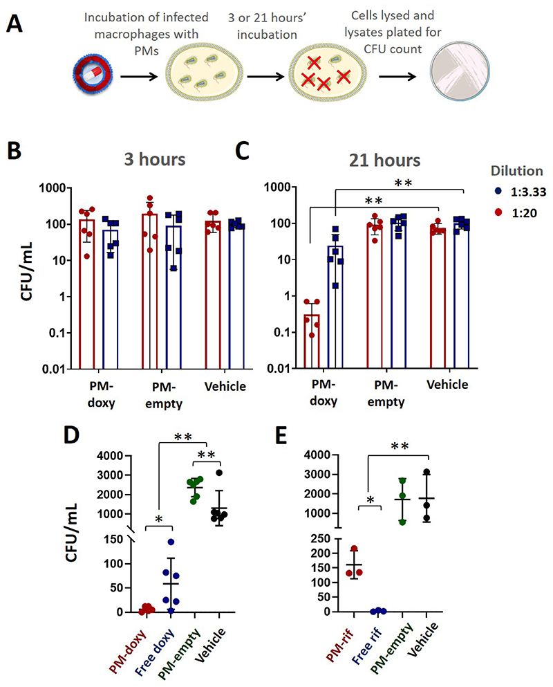
Melioidosis caused by the facultative intracellular pathogen Burkholderia pseudomallei is difficult to treat due to poor intracellular bioavailability of antibiotics and antibiotic resistance. In the absence of novel compounds, polymersome (PM) encapsulation may increase the efficacy of existing antibiotics and reduce antibiotic resistance by promoting targeted, infection-specific intracellular uptake. In this study, we developed PMs composed of widely available poly(ethylene oxide)-polycaprolactone block copolymers and demonstrated their delivery to intracellular B. thailandensis infection using multispectral imaging flow cytometry (IFC) and coherent anti-Stokes Raman scattering microscopy. Antibiotics were tightly sequestered in PMs and did not inhibit the growth of free-living B. thailandensis. However, on uptake of antibiotic-loaded PMs by infected macrophages, IFC demonstrated PM colocalization with intracellular B. thailandensis and a significant inhibition of their growth. We conclude that PMs are a viable approach for the targeted antibiotic treatment of persistent intracellular Burkholderia infection.

Keywords: CARS imaging; Raman spectroscopy; antibiotics; imaging flow cytometry; intracellular bacteria; nanoparticles; polymersomes.


Conflict of interest statement

Associated Content

The authors confirm that they have no competing financial interests.

Figures

Figure 1
Figure 1. Encapsulation and retention of doxycycline and rifampicin by PMs.
(A) Model for the different PM loading strategies for the antibiotics rifampicin and doxycycline. (B) The absorption spectrum of free doxycycline is similar to that of its spectrum when loaded into PMs after subtraction of unloaded PMs (C) The same is true of rifampicin. (D) A schematic of the assay used to assess PM retention of the antibiotic after a 14-day period. Samples were left to dialyse for 14 days, with regular antibiotic concentration reading time points. (E) After initial burst release, PM-doxycycline maintained an encapsulated concentration of 15.2 μg/ml ± 2.2 μg/ml and PM-rifampicin 8.5 μg/ml ± 2.4 μg/ml. Data points plotted show the mean of three formulations, n = 3. Graph transformed using one phase decay analysis. (F) DLS data displaying the PM-antibiotic hydrodynamic radius distributions over 14 days. (G) The size and PdI values of PM-doxycycline and PM-rifampicin over a 14-day period. DLS data based on measurements from one batch of each type of PMs, performed in three technical repeats.
Figure 2
Figure 2. Uptake of PMs by RAW 264.7 macrophage cells using label-free imaging.
(A) After incubation with DiI-labelled PMs, RAW 264.7 cells show cytoplasmic fluorescent puncta. (B) The Raman spectra for both PMs and the PEO-PCL polymer. A characteristic peak is present at approximately 2800 cm-1 indicating C-H bonds (C) CARS label-free imaging showing a comparison of untreated RAW 264.7 cells versus RAW 264.7 cells exposed to PM-empty nanoparticles. (D) PM-empty treated RAW 264.7 cells showed a significantly higher mean number of CARS spots per μm compared to the untreated control, p < 0.0001. CARS quantitative analysis data shows the mean and SD of one biological repeat performed in triplicate. (E) DiI fluorescent puncta in RAW 264.7 cells co-localise with lysosomes labelled with Lysotracker.
Figure 3
Figure 3. Intracellular infection of macrophages by B. thailandensis is heterogeneous and increased with time.
(A) Confocal imaging shows GFP-expressing B. thailandensis are present within RAW 264.7 cells after 3 hours incubation in the presence of kanamycin. Scale bar = 20 μm (B) Gating strategy for separating populations of B. thailandensis-positive (R5) and negative (R9) cells. (C) Gating strategy for separating RAW 264.7 cells with intracellular (R6) or extracellular (R14) B. thailandensis. (D) Representative ImageStream images of RAW264.7 cells with no bacteria present (R9), extracellular bacteria (R14) or intracellular bacteria (R6). Scale bar = 7 μM (E) There is a decrease in the percentage of cells with no bacteria present and an increase in those with either intracellular or extracellular bacteria between 3 and 21 hours.
Figure 4
Figure 4. Imaging flow cytometry analysis of PM-DiD uptake by RAW 264.7 macrophage cells.
(A) Fluorescent DiD puncta are visible in RAW 264.7 cells infected with GFP-labelled B. thailandensis. (B) Almost all RAW 264.7 cells are positive for DiD-labelled polymersomes after 3 hours (C) In bacteria-exposed populations, cells which contain intracellular bacteria take up the most PMs and those with no bacteria present take up the least, as measured by DiD fluorescence intensity. All exposed populations take up significant fewer PMs compared to unexposed macrophages. (* indicates p<0.01 and ** <0.001; data reflect means of 6 independent experiments). (D) Representative ImageStream images from the populations shown in (C). (E) Fluorescent puncta of DiD labelled PMs (Ch11) partially colocalise with GFP-positive bacteria, as shown by representative ImageStream images. (F) Gating strategy to separate RAW 264.7 cells with high co-localisation compared to low co-localisation. (G) The degree of PM/bacterial colocalisation declined from 3 hours to 24 hours. All ImageStream data is taken from three biological repeats, n=3, each performed in duplicate. ** = p < 0.0001.
Figure 5
Figure 5. PM-antibiotic preparations do not inhibit growth of free B. thailandensis.
(A) Both unloaded antibiotics were effective at inhibiting growth at the higher concentrations tested, at the 24-hour time point. Data is presented as the mean and SEM of one biological repeat tested in triplicate, n=1. (B) PM-rifampicin preparations and PM-doxycycline preparations do not inhibit the growth of B. thailandensis growing free in L-broth culture, at any concentration tested. Data is presented as the mean and SEM of one biological repeat tested in triplicate.
Figure 6
Figure 6. PM-antibiotic preparations reduce intracellular B. thailandensis burden.
(A) A schematic showing the assay used to assess intracellular killing. (B) After a 3-hour incubation of infected cells with PM-doxycycline there was no inhibition of intracellular growth observed. (C) After a 21-hour incubation, there was a significant level of intracellular bacterial killing. Data is presented as the mean and SD of data from two independent experiments, n=2, performed in triplicate and normalised to the 1:20 control group. (D) PM-doxycycline (1:3.33 dilution) significantly reduced intracellular bacteria burden compared to free doxycycline (4.5 μg/mL; p <0.05), unloaded PMs or vehicle controls (p <0.01) n=6; two independent experiments, mean and SD shown. (E) PM-rifampicin significantly reduced intracellular bacteria burden compared to unloaded PMs or vehicle controls (p <0.01). Free rifampicin (2.6 μg/ml) reduced intracellular bacterial burden compared to PM-rifampicin, PM-empty or vehicle controls n=3, mean and SD shown.

References

    1. Limmathurotsakul D, Peacock SJ. Melioidosis: A Clinical Overview. British Medical Bulletin. 2011;99(1):125–139. doi: 10.1093/bmb/ldr007. - DOI - PubMed
    1. Limmathurotsakul D, Golding N, Dance DA, Messina JP, Pigott DM, Moyes CL, Rolim DB, Bertherat E, Day NP, Peacock SJ, Hay SI. Predicted Global Distribution of Burkholderia pseudomallei and Burden of Melioidosis. Nat Microbiol. 2016;1(1) doi: 10.1038/nmicrobiol.2015.8. - DOI - PubMed
    1. Titball RW, Burtnick MN, Bancroft GJ, Brett P. Burkholderia pseudomallei and Burkholderia mallei Vaccines: Are We Close to Clinical Trials? Vaccine. 2017;35(44):5981–5989. doi: 10.1016/j.vaccine.2017.03.022. - DOI - PubMed
    1. Gilad J, Harary I, Dushnitsky T, Schwartz D, Amsalem Y. Burkholderia mallei and Burkholderia pseudomallei as Bioterrorism Agents: National Aspects of Emergency Preparedness. Israel Medical Association Journal. 2007;9(7):499–503. - PubMed
    1. Willcocks SJ, Denman CC, Atkins HS, Wren BW. Intracellular Replication of the Well-Armed Pathogen Burkholderia pseudomallei . Current Opinion in Microbiology. 2016;29:94–103. doi: 10.1016/i.mib.2015.11.007. - DOI - PubMed

Publication types

Substances

Supplementary concepts