. 2021 Jun 25;372(6549):eabf6548.
doi: 10.1126/science.abf6548. Epub 2021 Aug 5.

Ubiquitination of G3BP1 mediates stress granule disassembly in a context-specific manner

Affiliations

Ubiquitination of G3BP1 mediates stress granule disassembly in a context-specific manner

Youngdae Gwon et al. Science. .

Abstract

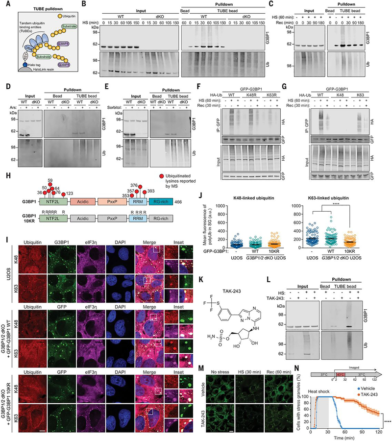
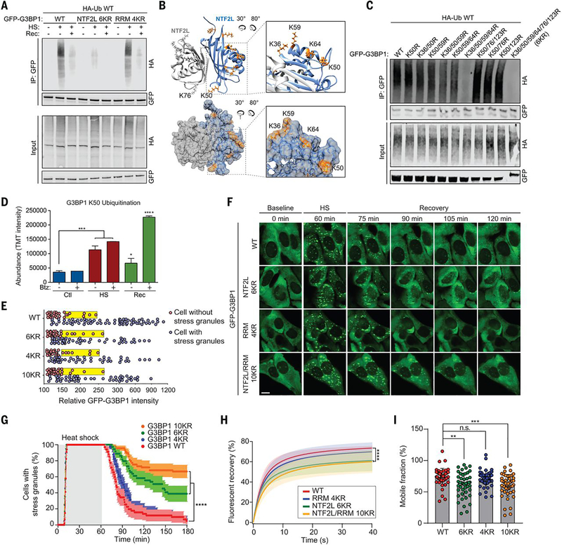
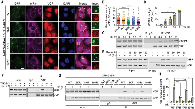
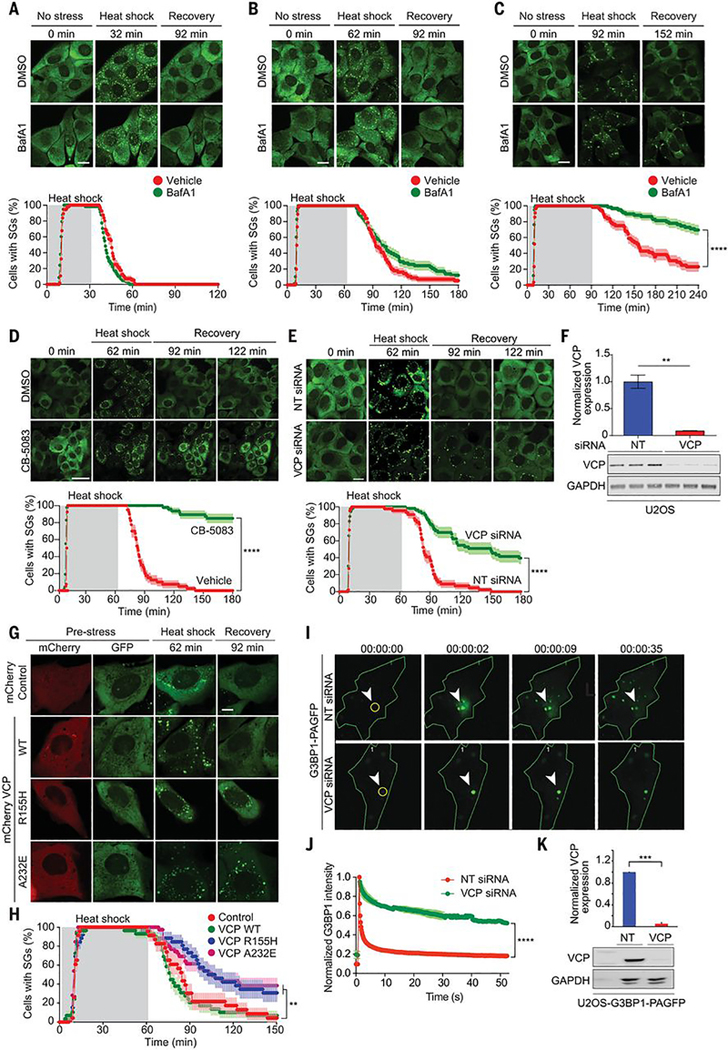
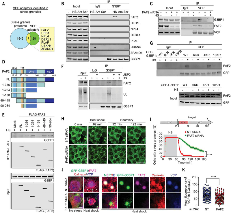
Stress granules are dynamic, reversible condensates composed of RNA and protein that assemble in eukaryotic cells in response to a variety of stressors and are normally disassembled after stress is removed. The composition and assembly of stress granules is well understood, but little is known about the mechanisms that govern disassembly. Impaired disassembly has been implicated in some diseases including amyotrophic lateral sclerosis, frontotemporal dementia, and multisystem proteinopathy. Using cultured human cells, we found that stress granule disassembly was context-dependent: Specifically in the setting of heat shock, disassembly required ubiquitination of G3BP1, the central protein within the stress granule RNA-protein network. We found that ubiquitinated G3BP1 interacted with the endoplasmic reticulum–associated protein FAF2, which engaged the ubiquitin-dependent segregase p97/VCP (valosin-containing protein). Thus, targeting of G3BP1 weakened the stress granule–specific interaction network, resulting in granule disassembly.


Conflict of interest statement

Competing interests: J.P.T. is a consultant for 5AM and Third Rock Ventures.

Figures

Fig. 1.
Fig. 1.. G3BP1 undergoes K63-linked ubiquitination in response to heat stress.
(A) Illustration showing TUBE capture of ubiquitinated G3BP1. TEV, tobacco etch virus protease cleavage site. (B and C) Immunoblots of TUBE-captured cell extracts showing levels of ubiquitinated G3BP1 in U2OS cells after different durations of 43°C heat shock, using U2OS G3BP1/2 dKO cells as controls (B), and during 37°C recovery (C). (D and E) Immunoblots of TUBE-captured cell extracts showing levels of ubiquitinated G3BP1 in response to oxidative stress (0.5 mM NaAsO2, 1 hour) (D) or osmotic stress (0.4 M sorbitol, 1 hour) (E). (F and G) Immunoblots of cell extracts captured with antibody to GFP, showing K63-linked ubiquitination of G3BP1. Transfected HEK293T cells were exposed to heat shock (43°C, 1 hour) and recovery (37°C, 30 min). K48R and K63R prevent the formation of K48-linked (K48R) or K63-linked (K63R) chains; K48 and K63 permit K48-linked or K63-linked chains exclusively. HA, hemagglutinin; IP, immunoprecipitate. (H) G3BP1 domain labeled with lysines on which ubiquitination has been reported. In the G3BP1 10KR mutant, six lysines in NTF2L and four lysines in RRM are mutated to arginine. (I) Immunofluorescent staining of fixed U2OS WT and U2OS G3BP1/2 dKO cells stably expressing GFP-G3BP WT and 10KR. Scale bar, 10 μm. (J) Fluorescence intensities of K48- and K63-linked ubiquitin in eIF3η-positive stress granules from three technical replicates are plotted in (n = 90). Error bars indicate SEM. ****P < 0.0001 (ANOVA with Tukey’s test). (K) Structure of TAK-243. (L) Immunoblot of TUBE-captured cell extracts showing block of heat shock–induced G3BP1 ubiquitination by TAK-243. U2OS cells were treated with DMSO or TAK-243 for 1 hour prior to heat shock. (M and N) Fluorescent imaging of U2OS cells stably expressing GFP-G3BP1 were treated with DMSO or TAK-243 (1 hour) prior to imaging. Representative images are shown in (M). In (N), GFP signals were monitored at 30-s intervals to count cells with two or more stress granules from three technical replicates (vehicle n = 32, TAK-243 n = 35). Scale bar, 20 μm. Error bars indicate SEM. ****P < 0.0001 (Mantel-Cox test).
Fig. 2.
Fig. 2.. Ubiquitination of the NTF2L domain of G3BP1 is required for the disassembly of stress granules.
(A) Immunoblot of transfected HEK293T cell extracts captured with antibody to GFP after exposure to no stress, heat shock for 1 hour, or 37°C recovery for 30 min. (B) Dimeric structure of G3BP1 NTF2L domain. K36, K50, K59, and K64 are highlighted in enlarged images (orange). (C) Immunoblot of transfected HEK293T cell extract captured with GFP antibody after exposure to heat shock for 1 hour. (D) TMT intensity of ubiquitinated G3BP1 peptides during heat shock and recovery with or without bortezomib (Btz). Error bars indicate SD. *P < 0.05, ***P < 0.001, ****P < 0.0001 (ANOVA with Dunnett’s test). (E) Percolation threshold of GFP-G3BP1 WT, 6KR, 4KR, and 10KR exposed to heat shock for 1 hour. U2OS G3BP1/2 dKO cells without stress granules (red) and with stress granules (blue) are plotted from three biological replicates according to GFP intensities (WT n = 109, 6KR n = 86, 4KR n = 78, 10KR n = 85). Yellow boxes indicate the cells with 25% highest levels of GFP within the stress granule–null group. (F) Stress granule disassembly in U2OS G3BP1/2 dKO cells expressing G3BP1 WT, 6KR, 4KR, or 10KR. Scale bar, 20 μm. (G) GFP signals were monitored at 30-s intervals at 37°C for 2 min, 43°C for 60 min, and 37°C for 118 min to count cells with two or more stress granules from three biological replicates (WT n = 38, 6KR n = 36, 4KR n = 39, 10KR n = 39). Error bars indicate SEM. ****P < 0.0001 (Mantel-Cox test). (H) FRAP of GFP-positive puncta in U2OS G3BP1/2 dKO cells transfected with GFP-G3BP1 WT, 6KR, 4KR, or 10KR and exposed to heat shock (1 hour) followed by recovery (10 min) from three biological replicates is plotted as mean ± SEM (WT n = 48, 6KR n = 48, 4KR n = 45, 10KR n = 49). ****P < 0.0001 (ANOVA with Tukey’s test). (I) Quantification of mobile fraction at 40 s of recovery from three biological replicates (WT n = 48, 6KR n = 48, 4KR n = 45, 10KR n = 49). Error bars indicate SD. **P < 0.01, ***P < 0.001 (ANOVA with Dunnett’s test); n.s., not significant.
Fig. 3.
Fig. 3.. VCP interacts with ubiquitinated G3BP1 in response to heat shock.
(A) Fluorescent imaging of U2OS G3BP1/2 dKO cells stably expressing GFP-G3BP WT, 6KR, 4KR or 10KR exposed to heat shock for 1 hour. Scale bar, 20 μm. (B) Fluorescence intensities of VCP in eIF3η-positive stress granules from three technical replicates (n = 90). Error bars indicate SEM. ****P < 0.0001 (ANOVA with Tukey’s test). (C) Immunoblot of U2OS cell extracts exposed to heat shock for the indicated times. Cell extracts were captured with magnetic beads conjugated with VCP antibody for IP, and resulting beads were analyzed by immunoblot. (D) Quantification of immunoblots from three biological replicates. Error bars indicate SEM. **P < 0.01, ***P < 0.001 (ANOVA with Tukey’s test). (E) Immunoblot showing G3BP1-VCP interaction in U2OS cells based on duration of recovery after heat shock for 2 hours. Cell extracts were captured with magnetic beads conjugated with antibody to VCP for IP, and the resulting beads were analyzed by immunoblot. (F) Immunoblot of U2OS cells treated with DMSO or TAK-243 (60 min) and exposed to heat shock for 2 hours. Cell extracts were captured with magnetic beads conjugated with antibody to VCP for IP. Blots show an attenuated G3BP1-VCP interaction with inhibition of ubiquitination. (G) Immunoblot of U2OS G3BP1/2 dKO cells stably expressing G3BP1 WT, 6KR, 4KR, or 10KR and exposed to heat shock for 2 hours. Cell extracts were captured with magnetic beads conjugated with antibody to GFP for IP. (H) Quantification of immunoblots from three biological replicates. Error bars indicate SEM. ***P < 0.001 (ANOVA with Tukey’s test).
Fig. 4.
Fig. 4.. VCP regulates disassembly of stress granules.
(A to C) Top: U2OS/GFP-G3BP1 were treated with DMSO or bafilomycin A1 (BafA1) for 18 hours before live imaging. Scale bar, 20 μm. Bottom: GFP signals were monitored at 30-s intervals at 37°C for 2 min, 43°C for 30 min (A), 60 min (B), or 90 min (C), and 37°C for 88 min (A), 118 min (B), or 148 min (C) to count cells with two or more stress granules from three technical replicates [DMSO n = 56, BafA1 n = 58 in (A); DMSO n = 56, BafA1 n = 58 in (B); DMSO n = 52, BafA1 n = 59 in (C)]. Error bars indicate SEM. ****P < 0.0001 (Mantel-Cox test). (D and E) U2OS/GFP-G3BP1 cells were treated with DMSO or CB-5083 (1 hour) (D) or transfected with nontargeting (NT) or VCP siRNA (E) before live imaging. GFP signals were monitored at 30-s intervals at 37°C for 2 min, 43°C for 60 min, and 37°C for 118 min to count the cells with two or more stress granules [DMSO n = 40, CB-5083 n = 47 in (D); NT siRNA n = 44, VCP siRNA n = 53 in (E)]. Scale bars, 50 μm (D), 20 μm (E). Error bars indicate SEM. ****P < 0.0001 (Mantel-Cox test). (F) Immunoblot of U2OS cells transfected with NT or VCP siRNA from three biological replicates. Error bars indicate SEM. **P < 0.01 (Student’s t test). (G) U2OS/GFP-G3BP1 cells were transfected with pmCherry-N1, VCP WT-mCherry, VCP R155H-mCherry, or VCP A232EmCherry. Scale bar, 10 μm. (H) GFP signals of mCherry-positive cells were monitored at 60-s intervals at 37°C for 2 min, 43°C for 60 min, and 37°C for 88 min to count cells with two or more stress granules from three biological replicates (control n = 23, VCP WT n = 29, VCP R155H n = 26, VCP A232E n = 39). Error bars indicate SEM. **P < 0.01 (Mantel-Cox test). (I) U2OS cells expressing photoactivatable G3BP1 (G3BP1-PAGFP) were transfected with NT or VCP siRNA. (J) Intensities of GFP signals within activated ROIs were monitored at 200-ms intervals from three technical replicates. ****P < 0.0001 (ANOVA with Sidak’s test). (K) Immunoblots of U2OS cells expressing G3BP1-PAGFP transfected with NT or VCP siRNA from three biological replicates. ***P< 0.001 (Student’s t-test).
Fig. 5.
Fig. 5.. FAF2 links ubiquitinated G3BP1 to VCP.
(A) Venn diagram showing overlapping proteins among the stress granule proteome and known VCP adaptors. (B) Immunoblot of U2OS cells extracts capture with antibody to GFP exposed to no stress, heat shock (1.5 hours), oxidative stress (sodium arsenite, 1.5 hours), or osmotic stress (sorbitol, 1.5 hours). (C) Immunoblot of U2OS cells extracts captured with antibody to VCP after transfection with NT or FAF2 siRNA and exposure to heat shock for 1.5 hours. (D) Domain structure of human FAF2 protein and deletion constructs used to investigate the function of individual domains of FAF2. UBA, ubiquitin-associated domain; TM, transmembrane domain; UAS, upstream activation sequence domain; UBX, ubiquitin regulatory X domain. (E) Immunoblot of U2OS cells captured with antibody to FLAG exposed to heat shock for 1.5 hours after transfection of FLAG-FAF2 full length (FL) or deletion mutants. (F) Immunoblot of U2OS cells exposed to no stress or heat shock for 1.5 hours. Cell extracts were incubated with or without purified USP2 and captured with magnetic beads conjugated with antibody to G3BP1 for IP, and the resulting beads were analyzed by immunoblot. (G) Immunoblots of U2OS G3BP1/2 dKO cells stably expressing G3BP1 WT, 6KR, 4KR, or 10KR mutants and exposed to heat shock for 2 hours. Cell extracts were captured with magnetic beads conjugated with antibody to GFP for IP. (H) U2OS/GFP-G3BP1 cells were transfected with NT or FAF2 siRNA. Scale bar, 50 μm. (I) GFP signals were monitored at 30-s intervals at 37°C for 2 min, 43°C for 60 min, and 37°C for 118 min to count cells with two or more stress granules from three technical replicates (NT siRNA n = 47, FAF siRNA n = 59). Error bars indicate SEM. ****P < 0.0001 (Mantel-Cox test). (J) Fluorescent imaging of U2OS/GFP-G3BP1 cells exposed to heat shock for 1 hour. Scale bar, 10 μm. (K) Fluorescence intensities of VCP in stress granules from three biological replicates are plotted as mean ± SEM (NT siRNA n = 272, FAF2 siRNA n = 349). ****P < 0.0001 (Student’s t test).

Comment in

References

    1. Buchan JR, mRNP granules. Assembly, function, and connections with disease. RNA Biol. 11, 1019–1030 (2014). - PMC - PubMed
    1. Gomes E, Shorter J, The molecular language of membraneless organelles. J. Biol. Chem 294, 7115–7127 (2019). - PMC - PubMed
    1. Yang P et al., G3BP1 Is a Tunable Switch that Triggers Phase Separation to Assemble Stress Granules. Cell 181, 325–345 e328 (2020). - PMC - PubMed
    1. Sanders DW et al., Competing Protein-RNA Interaction Networks Control Multiphase Intracellular Organization. Cell 181, 306–324 e328 (2020). - PMC - PubMed
    1. Guillen-Boixet J et al., RNA-Induced Conformational Switching and Clustering of G3BP Drive Stress Granule Assembly by Condensation. Cell 181, 346–361 e317 (2020). - PMC - PubMed

Publication types

MeSH terms