

Ocular abnormalities in Polish Hunting Dogs
- PMID: 34739488
- PMCID: PMC8570502
- DOI: 10.1371/journal.pone.0258636
Ocular abnormalities in Polish Hunting Dogs
Abstract
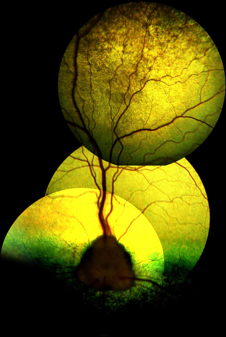
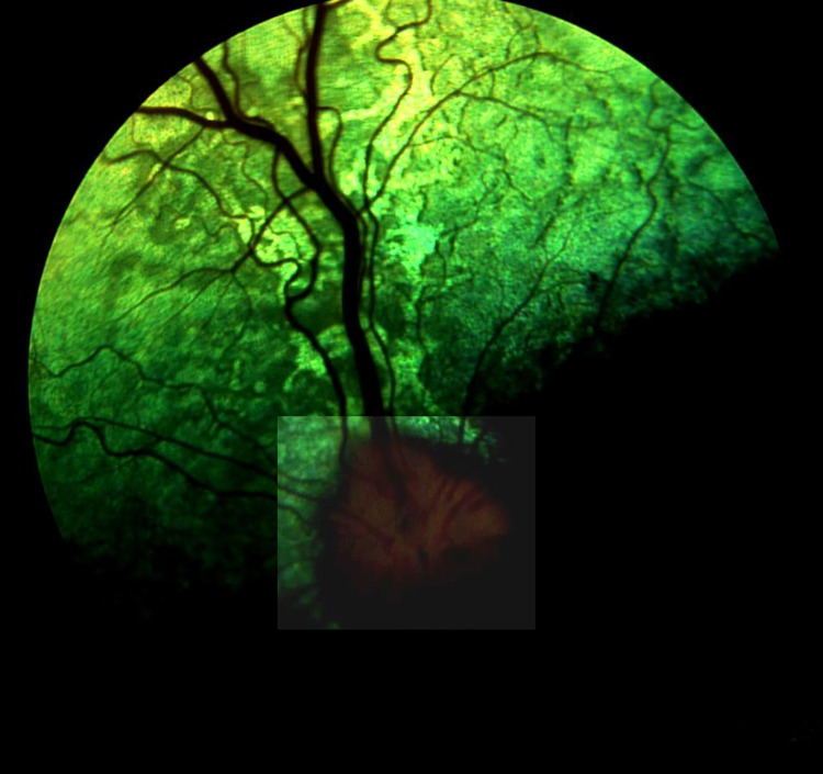
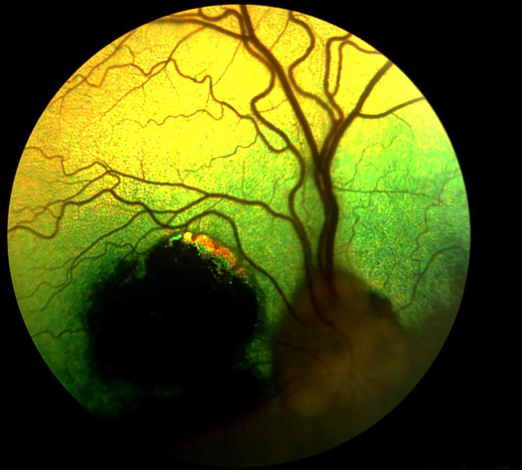
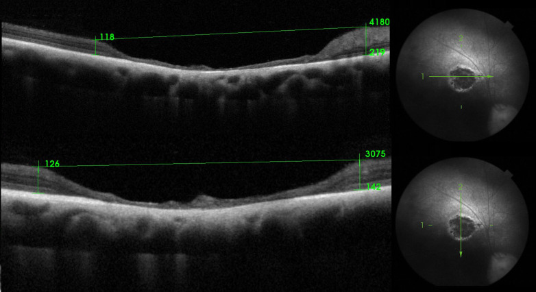
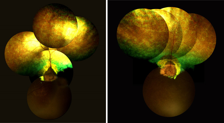
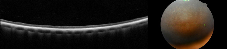
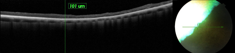
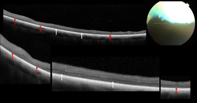
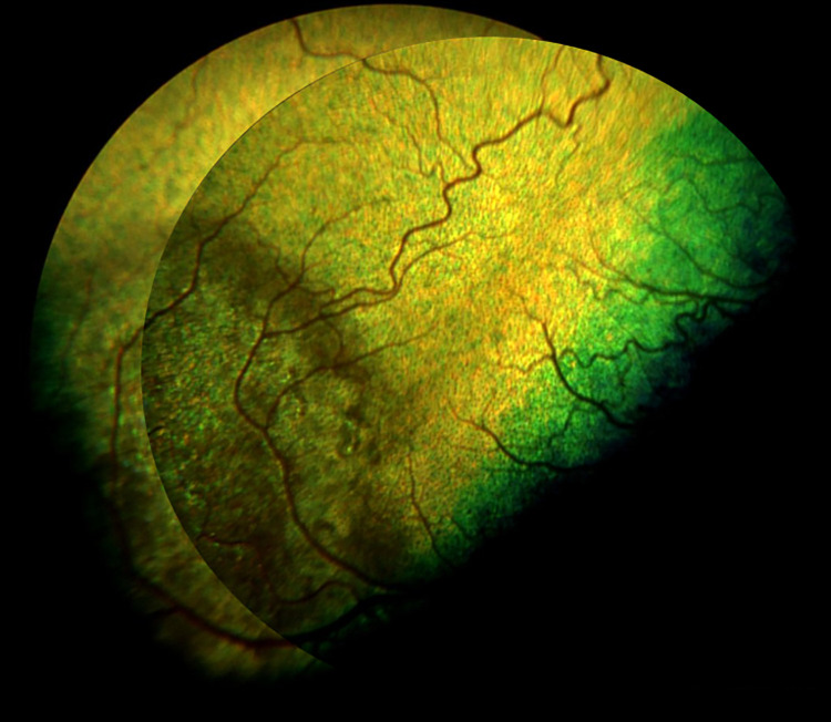
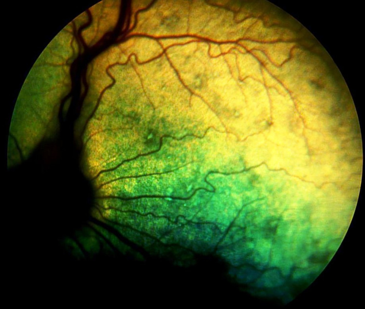
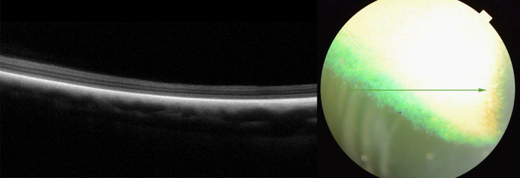
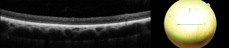
This study aimed to describe and determine the prevalence of ocular abnormalities in Polish Hunting Dogs. The study was conducted with 193 Polish Hunting Dogs: 101 female and 92 male animals, aged between 3 months and 12 years. Ophthalmic examinations were performed using slit lamp biomicroscopy, ophthalmoscopy, and tonometry based on the ophthalmological protocol for the examination of hereditary eye diseases. Spectral-domain optical coherence tomography (SD-OCT) was performed for dogs with sudden acquired retinal degeneration syndrome (SARDS) and progressive retinal atrophy (PRA), while electroretinography was also performed in dogs with SARDS. Five dogs (2.6%) were diagnosed with cataract, iris coloboma in 3 dogs (1.6%), ocular dermoid in 1 dog (0.5%), and retinal dysplasia, distichiasis and entropion in 1 dog (1%). Three dogs (1.6%) were diagnosed with PRA and SARDS occurred in 1 dog. Retinal lesions was observed in 16 dogs (8.3%). The clinical signs of retinopathy observed in Polish Hunting Dogs included discoloration of the tapetal fundus, patchy increased reflectivity in the region of discoloration, focus of hyperpigmentation and an area of tapetal hyper-reflectivity with a pigmented center. SD-OCT performed in the 3 dogs with PRA revealed alteration in the retinal layers, which was most advanced in the non-tapetal fundus. Although SD-OCT revealed retinal layers with normal architecture only in some parts of the dorsal, nasal and temporal regions in dogs with SARDS, areas of disorganized external limiting membrane, myeloid zone, ellipsoid zone, outer photoreceptor segment and interdigitation zone were also observed. Polish Hunting Dogs should undergo periodic ophthalmological examination for the evaluation of other hereditary eye diseases. The prevalence of retinal lesions in Polish Hunting Dogs requires further research.
Conflict of interest statement
The authors have declared that no competing interests exist.
Figures
Similar articles
-
Evaluation of retinal morphology of canine sudden acquired retinal degeneration syndrome using optical coherence tomography and fluorescein angiography.Vet Ophthalmol. 2019 Jul;22(4):398-406. doi: 10.1111/vop.12602. Epub 2018 Aug 22. Vet Ophthalmol. 2019. PMID: 30136357
-
Morphometric assessment of the choroid in dogs diagnosed with retinal atrophy (RA) with symptoms of progressive retinal atrophy, using spectral-domain optical coherence tomography (SD-OCT).Pol J Vet Sci. 2024 Jun;27(2):261-270. doi: 10.24425/pjvs.2024.149356. Pol J Vet Sci. 2024. PMID: 39736068
-
Optical coherence tomography and molecular analysis of sudden acquired retinal degeneration syndrome (SARDS) eyes suggests the immune-mediated nature of retinal damage.Vet Ophthalmol. 2019 May;22(3):305-327. doi: 10.1111/vop.12597. Epub 2018 Aug 15. Vet Ophthalmol. 2019. PMID: 30109754 Free PMC article.
-
Multimodal fundus imaging in fundus albipunctatus with RDH5 mutation: a newly identified compound heterozygous mutation and review of the literature.Doc Ophthalmol. 2012 Aug;125(1):51-62. doi: 10.1007/s10633-012-9336-z. Epub 2012 Jun 6. Doc Ophthalmol. 2012. PMID: 22669287 Review.
-
Is it canine DUSN?: Another view of retinopathies, some acquired, and others possibly "inherited": Another view of retinopathies, some acquired, and others possibly "inherited".Vet Ophthalmol. 2022 Mar;25(2):96-108. doi: 10.1111/vop.12951. Epub 2021 Dec 11. Vet Ophthalmol. 2022. PMID: 34894198 Free PMC article. Review.
Cited by
-
Analysis of Selected Eye Disorders in a Group of Predisposed Breeds of Dogs: Molecular Diagnostics of Collie Eye Anomaly and Progressive Retinal Atrophy.Genes (Basel). 2025 Apr 23;16(5):474. doi: 10.3390/genes16050474. Genes (Basel). 2025. PMID: 40428296 Free PMC article.
References
-
- Brabletz A. Gończy Polski. PIES—Dwumiesięcznik ZKwP—special edition. 2006;61–69.
-
- Kaźmierski A. Ogary i Gończe polskie. ZKwP, Opole; 2014. pp. 20149–10.
-
- Goleman M, Balicki I, Radko A, Jakubczak A, Fornal A. Genetic diversity of the Polish Hunting Dog population based on pedigree analyses and molecular studies. Livest. Sci. 2019; 229: 114–117. doi: 10.1016/j.livsci.2019.09.017 - DOI
-
- Genetics Committee of the American College of Veterinary Ophthalmologists. Ocular Disorders Presumed to be Inherited in Purebred Dogs (“The Blue Book”). 2019. [cited 2021 April 27]. Available from: https://www.ofa.org/wp-content/uploads/2021/01/Bluebook-V12.pdf
-
- Narfstrom K, Peterson-Jones S. Diseases of the canine ocular fundus. In: Gelatt KN, editors. Veterinary Ophthalmology, 4th ed. Blackwell Publishing, Ames; 2007. pp. 989–1000.
MeSH terms
LinkOut - more resources
Full Text Sources
Research Materials
