. 2021 Nov 5;21(1):1181.
doi: 10.1186/s12885-021-08933-y.

NOX4-derived ROS-induced overexpression of FOXM1 regulates aerobic glycolysis in glioblastoma

Affiliations

NOX4-derived ROS-induced overexpression of FOXM1 regulates aerobic glycolysis in glioblastoma

Xiangsheng Su et al. BMC Cancer. .

Abstract

Background: Increased expression of the transcription factor Forkhead box M1 (FOXM1) has been reported to play an important role in the progression and development of multiple tumors, but the molecular mechanisms that regulate FOXM1 expression remain unknown, and the role of FOXM1 in aerobic glycolysis is still not clear.

Methods: The expression of FOXM1 and NADPH oxidase 4 (NOX4) in normal brain tissues and glioma was detected in data from the TCGA database and in our specimens. The effect of NOX4 on the expression of FOXM1 was determined by Western blot, qPCR, reactive oxygen species (ROS) production assays, and luciferase assays. The functions of NOX4 and FOXM1 in aerobic glycolysis in glioblastoma cells were determined by a series of experiments, such as Western blot, extracellular acidification rate (ECAR), lactate production, and intracellular ATP level assays. A xenograft mouse model was established to test our findings in vivo.

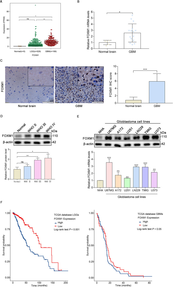
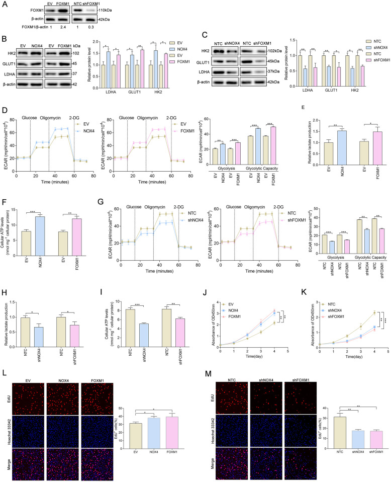
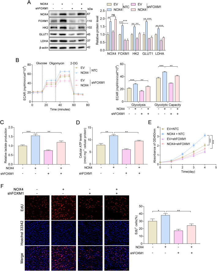
Results: The expression of FOXM1 and NOX4 was increased in glioma specimens compared with normal brain tissues and correlated with poor clinical outcomes. Aberrant mitochondrial reactive oxygen species (ROS) generation of NOX4 induced FOXM1 expression. Mechanistic studies demonstrated that NOX4-derived MitoROS exert their regulatory role on FOXM1 by mediating hypoxia-inducible factor 1α (HIF-1α) stabilization. Further research showed that NOX4-derived MitoROS-induced HIF-1α directly activates the transcription of FOXM1 and results in increased FOXM1 expression. Overexpression of NOX4 or FOXM1 promoted aerobic glycolysis, whereas knockdown of NOX4 or FOXM1 significantly suppressed aerobic glycolysis, in glioblastoma cells. NOX4-induced aerobic glycolysis was dependent on elevated FOXM1 expression, as FOXM1 knockdown abolished NOX4-induced aerobic glycolysis in glioblastoma cells both in vitro and in vivo.

Conclusion: Increased expression of FOXM1 induced by NOX4-derived MitoROS plays a pivotal role in aerobic glycolysis, and our findings suggest that inhibition of NOX4-FOXM1 signaling may present a potential therapeutic target for glioblastoma treatment.

Keywords: Aerobic glycolysis; FOXM1; Glioblastoma; NOX4; ROS.


Conflict of interest statement

The authors declare that they have no conflicts of interest.

Figures

Fig. 1
Fig. 1
FOXM1 is high expressed in glioma. A Analysis of FOXM1 expression in normal brain and glioma tissues based on data from the TCGA database. B qPCR detection of FOXM1 mRNA expression in normal brain and glioma tissues from clinical specimens. C IHC analysis of FOXM1 protein expression in normal brain and GBM samples. Scale bars, 20 μm. D Western blot analysis of FOXM1 protein expression in normal brain and glioma with different grades. E Western blot and qPCR detection of FOXM1 expression in multiple glioblastoma cells and in normal human astroglia (NHA) cell lines. F The prognostic value of FOXM1 expression in LGG and GBM was analyzed in TCGA datasets. ns, not significant; *, P < 0.05; **, P < 0.01; ***, P < 0.001
Fig. 2
Fig. 2
NOX4 stimulates FOXM1 expression by increasing mitochondrial ROS. A Analysis of NOX4 expression in normal brain and glioma tissues in TCGA datasets. B The prognostic value of NOX4 expression in LGG and GBM was analyzed in TCGA datasets. C qPCR detection of NOX4 mRNA expression in normal brain and glioma clinical specimens. D IHC analysis of NOX4 protein expression in normal brain and GBM. Scale bars, 20 μm. E Western blot analysis of NOX4 and FOXM1 protein expression in normal brain and glioma tissues of different grades. F The correlation between NOX4 expression and FOXM1 expression in normal brain and glioma patients according to the TCGA and our clinical samples. G Western blot and qPCR analysis of FOXM1 protein and mRNA levels in control or NOX4-overexpressing glioblastoma cells. H MitoSOX staining showing the upregulation of NOX4 affected the production of ROS in glioblastoma cells. Scale bars, 50 μm. I Western blot and qPCR analysis of FOXM1 protein and mRNA levels in control or NOX4 knockdown glioblastoma cells. J MitoSOX staining showed that the downregulation of NOX4 affected the production of ROS in glioblastoma cells. Scale bars, 50 μm. K Western blot analysis showing the protein expression of FOXM1 in glioblastoma cells exposed to NAC at different concentrations for 24 h. L Western blot analysis showing the protein expression of FOXM1 in glioblastoma cells exposed to H2O2 at different concentrations for 24 h. M Western blot analysis showing the protein expression of FOXM1 in glioblastoma cells treated with Mito-Tempo (1 mM) in the control or NOX4-overexpressing cells. N Immunofluorescence staining showing the subcellular localization of NOX4 in glioblastoma cells; the merge in yellow shows colocalization. Scale bar 50 μM. *, P < 0.05; **, P < 0.01; ***, P < 0.001
Fig. 3
Fig. 3
NOX4 positively regulates FOXM1 expression by mediating HIF-1α stabilization. A Western blot analysis showing HIF-1α protein levels in NOX4-overexpressing or NOX4-knockdown glioblastoma cells. B Western blot analysis showing HIF-1α protein levels in NOX4-knockdown glioblastoma cells. C qPCR detection of FOXM1 mRNA levels in HIF-1α-overexpressing glioblastoma cells. D Western blot analysis showing the FOXM1 protein levels in HIF-1α-overexpressing glioblastoma cells. E Western blot analysis showing the FOXM1 protein levels in normoxic and hypoxic glioblastoma cells. F The indicated glioblastoma cells were transfected with HIF-1α siRNA or negative control siRNA, and the protein levels of FOXM1, HIF-1α, and NOX4 were measured by Western blot. G Western blot analysis showing the protein levels of the FOXM1 downstream targets cyclin B1 and PLK1 in normoxic and hypoxic glioblastoma cells. H Map of the HIF-1α binding site sequence from the JASPAR database. I Sequences and positions of putative HIF-1α-binding regions on the FOXM1 promoter and luciferase reporter constructs for wild-type or mutant FOXM1 promoter were cotransfected into U87MG cells along with EV- or HIF-1α-overexpressing plasmids for 48 h. Promoter activity was examined with a dual-luciferase reporter assay kit. *, P < 0.05; **, P < 0.01; ***, P < 0.001
Fig. 4
Fig. 4
NOX4 and FOXM1 regulate aerobic glycolysis and proliferation in glioblastoma cells. A Western blot analysis showing the overexpression and silencing efficacy of FOXM1. B Western blot analysis showing that NOX4 or FOXM1 overexpression increased the protein levels of the glycolytic genes LDHA, GLUT1, and HK2. C Western blot analysis showing that NOX4 or FOXM1 knockdown decreased the protein levels of the glycolytic genes HK2, GLUT1, and LDHA. D ECAR was measured in NOX4- or FOXM1-overexpressing U87MG cells using an XFe96 Extracellular Flux Analyzer. Glycolysis and glycolytic capacity were increased in NOX4- or FOXM1-overexpressing cells. E-F NOX4 or FOXM1 overexpression increased lactate production and cellular ATP levels. G ECAR was measured in NOX4- or FOXM1-knockdown U87MG cells using an XFe96 Extracellular Flux Analyzer. Glycolysis and glycolytic capacity were decreased in NOX4- or FOXM1-knockdown cells. H-I NOX4 or FOXM1 knockdown decreased lactate production and cellular ATP levels. J-K The viability of U87MG cells was increased upon NOX4 or FOXM1 overexpression and decreased upon NOX4 or FOXM1 knockdown, as determined with CCK-8. L-M NOX4 or FOXM1 overexpression increased the proliferation rate of U87MG cells, whereas NOX4 or FOXM1 knockdown with specific shRNA decreased the proliferation rate of U87MG cells, as shown by EdU staining. *, P < 0.05; **, P < 0.01; ***, P < 0.001
Fig. 5
Fig. 5
FOXM1 is critical for NOX4-induced aerobic glycolysis and proliferation in glioblastoma cells. A Western blot analysis showing the protein expression of the indicated genes in U87MG cells. B U87MG cells were infected with NOX4 overexpression (NOX4) plasmid and FOXM1 knockdown (shFOXM1) lentiviral vectors before plating in a Seahorse XFe96 analyzer and assessment with a glycolysis stress test. C-D Overexpression of NOX4 promoted the production of lactate and cellular ATP, while knockdown of FOXM1 reversed these effects, in U87MG cells. E-F CCK-8 and EdU assays showing the proliferation of the indicated U87MG cells stably expressing NOX4 + shFOXM1. *, P < 0.05; **, P < 0.01; ***, P < 0.001
Fig. 6
Fig. 6
Altered NOX4-FOXM1 signaling modulates tumorigenesis in vivo. A H&E staining of the sections of xenograft mouse brains. Scale bar, 1 mm. B Tumor size (mm3) was measured. C-D IHC and Western blot analysis of the indicated proteins in tumor tissue samples from the indicated groups. Scale bar, 20 μm. E Cellular ATP levels were determined in tumors from xenograft mice. F Schematic model of NOX4-FOXM1 signaling-regulated aerobic glycolysis and progression in glioblastoma cells. In glioblastoma cells, NOX4-generated mitochondrial ROS mediate HIF-1α stabilization. Stabilized HIF-1α can directly bind to the FOXM1 promoter and subsequently promote the expression of FOXM1, resulting in aerobic glycolysis and progression. *, P < 0.05; **, P < 0.01; ***, P < 0.001

Similar articles

Cited by

References

    1. Tan AC, Ashley DM, Lopez GY, Malinzak M, Friedman HS, Khasraw M. Management of glioblastoma: state of the art and future directions. CA Cancer J Clin. 2020;70(4):299–312. doi: 10.3322/caac.21613. - DOI - PubMed
    1. Festuccia C, Biordi AL, Tombolini V, Hara A, Bailey D. Targeted molecular therapy in glioblastoma. J Oncol. 2020;2020:5104876–5104873. doi: 10.1155/2020/5104876. - DOI - PMC - PubMed
    1. Hanahan D, Weinberg RA. Hallmarks of cancer: the next generation. Cell. 2011;144(5):646–674. doi: 10.1016/j.cell.2011.02.013. - DOI - PubMed
    1. Liu Y, Cao Y, Zhang W, Bergmeier S, Qian Y, Akbar H, Colvin R, Ding J, Tong L, Wu S, Hines J, Chen X. A small-molecule inhibitor of glucose transporter 1 downregulates glycolysis, induces cell-cycle arrest, and inhibits cancer cell growth in vitro and in vivo. Mol Cancer Ther. 2012;11(8):1672–1682. doi: 10.1158/1535-7163.MCT-12-0131. - DOI - PubMed
    1. Pathria G, Scott DA, Feng Y, Sang Lee J, Fujita Y, Zhang G, et al. Targeting Glycolysis through Inhibition of Lactate Dehydrogenase Impairs Tumor Growth in Preclinical Models of Ewing Sarcoma. 2018;37(20). https://pubmed.ncbi.nlm.nih.gov/31431459/. - PMC - PubMed

MeSH terms