. 2021 Nov 5;20(1):142.
doi: 10.1186/s12943-021-01443-2.

ciRS-7 is a prognostic biomarker and potential gene therapy target for renal cell carcinoma

Affiliations

ciRS-7 is a prognostic biomarker and potential gene therapy target for renal cell carcinoma

Weipu Mao et al. Mol Cancer. .

Erratum in

Abstract

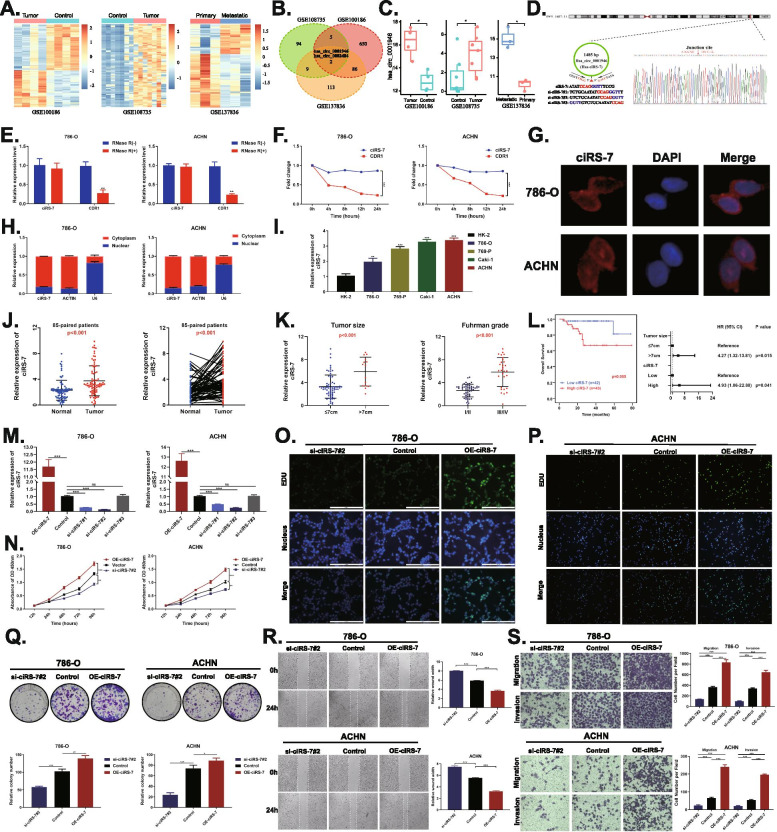
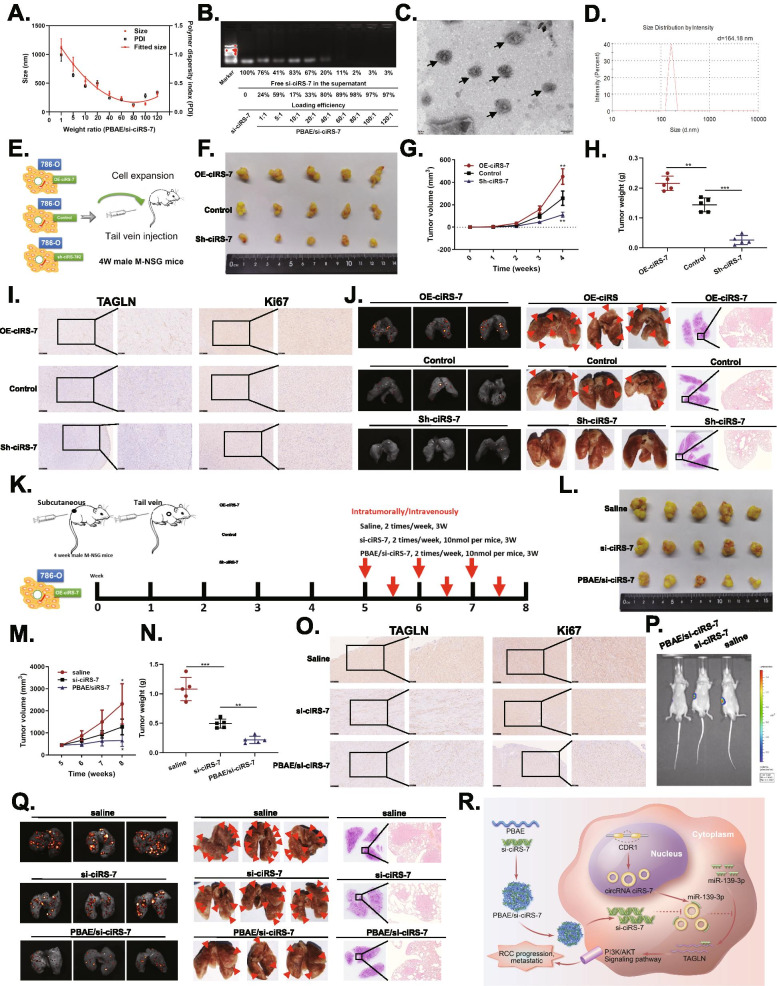
Circular RNAs are a new class of non-coding RNAs that have been shown to play critical roles in the development and progression of renal cell carcinoma (RCC). However, little is known about the functional mechanisms and therapeutic role of ciRS-7 in RCC. A series of in vitro and in vivo experiments were performed to investigate the functional mechanism and therapeutic role of ciRS-7, such as real-time quantitative PCR, CCK-8, wound healing, transwell, colony formation, Edu, tumor xenograft and lung metastasis in NSG mice. RNA pull-down, dual luciferase reporter, fluorescence in situ hybridization (FISH) and rescue assays were used to determine the relationship between ciRS-7, miR-139-3p and TAGLN. In addition, we constructed PBAE/si-ciRS-7 nanocomplexes with PBAE material to evaluate the therapeutic effect of the nanocomplexes on tumor in vivo. ciRS-7 was highly expressed in RCC tumor tissues and cell lines, and high ciRS-7 expression correlated with tumor size, high Fuhrman grade and poor survival. Depletion of ciRS-7 significantly inhibited RCC cell proliferation, invasion, tumor growth and metastasis in vivo, while overexpression of ciRS-7 had the opposite effect. Mechanistically, ciRS-7 acts as a "ceRNA" for miR-139-3p to prevent TAGLN degradation and promoting RCC progression and metastasis via the PI3K/AKT signaling pathway. In addition, miR-139-3p mimics or inhibitor could reverse the altered malignant tumor behavior caused by ciRS-7 overexpression or silencing. Furthermore, the PBAE/siciRS-7 nanocomplexes could significantly inhibit RCC tumor progression and metastasis in vivo. ciRS-7 acts as a tumor promoter by regulating the miR-139-3p/TAGLN axis and activating the PI3K/AKT signaling pathway to promote RCC progression and metastasis. Drug development of PBAE/si-ciRS-7 nanocomplexes targeting ciRS-7 may represent a promising gene therapeutic strategy for RCC.

Keywords: Gene therapeutic; Metastasis; PBAE/si-ciRS-7 nanocomplexes; Renal cell carcinoma; ciRS-7.


Conflict of interest statement

The authors declare that they have no competing interests.

Figures

Fig. 1
Fig. 1
ciRS-7 is overexpressed in RCC tissues and cells and promotes RCC cell proliferation, migration and invasion in vitro. A, Hierarchical clustering analysis of differentially expressed circRNAs in GSE100186, GSE108735 and GSE137836. B. Venn diagram of circRNAs commonly highly expressed in GSE100186, GSE108735 and GSE137836. C. Expression of ciRS-7 in GSE100186, GSE108735 and GSE137836. D. Sanger sequencing to verify the splice junctions of ciRS-7. E and F. qRT-PCR analysis of ciRS-7 and linear CDR1 in 786-O and ACHN cells treated with RNase R or actinomycin D. G. FISH assay to detect the cellular localization of ciRS-7. H. qRT-PCR analysis of ciRS-7 was conducted in nuclear and cytoplasmic fractions of 786-O and ACHN cells. I and J. ciRS-7 was highly expressed in RCC cell lines and tumour tissues. K. Relative expression levels of ciRS-7 in different tumor sizes and Fuhrman grade. L. Kaplan-Meier’s survival curves showed the correlations between ciRS-7 expression and OS, and multivariate Cox regression of hazard ratios for RCC OS. M. Relative expression of ciRS-7 was confirmed by qPCR in 786-O and ACHN cell lines transfected with OE-ciRS-7, control, si-ciRS-7#1, si-ciRS-7#2 or si-ciRS-7#3. N. Growth curves of 786-O and ACHN cell lines were measured after transfection with indicated vectors by CCK-8. O and P. Edu assay to detect cell proliferation capacity after transfection with the indicated vectors. Q, Colony formation assay to detect cell migration ability after transfection with the indicated vectors. R. Wound healing assay to detect cell migration ability after transfection with the indicated vectors. S. Transwell assay to detect cell migration and invasion ability after transfection with the indicated vectors. (*p < 0.05, **p < 0.01, ***p < 0.001)
Fig. 2
Fig. 2
ciRS-7 enhances RCC tumor growth and metastasis in vivo and PBAE/si-ciRS-7 nanocomplexes inhibits RCC growth and metastasis in vivo. A. The sizes with different weight ratios of PBAE to siPlk1. The weight ratios of PBAE to si-ciRS-7 were ranged from 1 to 120. B. The loading efficiency of PBAE. C. TEM images of PBAE/si-ciRS-7 nanocomplexes (PBAE/si-ciRS-7 = 80/1). D. size distribution of PBAE/si-ciRS-7 nanocomplexes (PBAE/si-ciRS-7 = 80/1). E. Graphic illustration of the in vivo mice model study. F-H. 786-O cells stably transfected with OE-ciRS-7, control, or sh-ciRS-7 were injected subcutaneously into the mice. Tumor volume (G) and weight (H) increased in the OE-ciRS-7 group, while both tumor volume and weight decreased in the sh-ciRS-7 group. I. IHC assay demonstrated the level of TAGLN and Ki67 in pairs of tumours. J. After tail vein injection of treated cells, imaging, gross lung tissue lesions and HE staining were observed. K. The flow diagram showed the scheme of intratumorally/intravenously with saline, si-ciRS-7 or PBAE/si-ciRS-7 into mice. L-N. Weight volume and weight change after 3 weeks of treatment with saline, si-ciRS-7 or PBAE/si-ciRS-7 for xenograft tumors or lung metastasis models. O. IHC assay demonstrated the level of TAGLN and Ki67 in pairs of tumours. P. IVIS imaging of mice treated with saline, si-ciRS-7 or PBAE/si-ciRS-7 for subcapsular orthotopic implantation of the right kidney. Q. After tail vein injection of treated cells, imaging, gross lung tissue lesions and HE staining were observed. R. The hypothetical model depicts the roles of ciRS-7 in the promotion of RCC. (*p < 0.05, **p < 0.01, ***p < 0.001)

References

    1. Siegel RL, Miller KD, Jemal A. Cancer statistics, 2020. CA Cancer J Clin. 2020;70(1):7–30. doi: 10.3322/caac.21590. - DOI - PubMed
    1. Lotan Y, Margulis V. Predicting recurrence in patients with localised renal cell carcinoma after nephrectomy. Lancet Oncol. 2019;20(4):473–475. doi: 10.1016/S1470-2045(19)30024-5. - DOI - PubMed
    1. Sung H, Ferlay J, Siegel RL, Laversanne M, Soerjomataram I, Jemal A, et al. Global Cancer statistics 2020: GLOBOCAN estimates of incidence and mortality worldwide for 36 cancers in 185 countries. CA Cancer J Clin. 2021;71(3):209–249. doi: 10.3322/caac.21660. - DOI - PubMed
    1. Capitanio U, Montorsi F. Renal cancer. Lancet. 2016;387(10021):894–906. doi: 10.1016/S0140-6736(15)00046-X. - DOI - PubMed
    1. Salzman J, Chen RE, Olsen MN, Wang PL, Brown PO. Cell-type specific features of circular RNA expression. PLoS Genet. 2013;9(9):e1003777. doi: 10.1371/journal.pgen.1003777. - DOI - PMC - PubMed

Publication types

MeSH terms