. 2022 Feb 1;411(1):112906.
doi: 10.1016/j.yexcr.2021.112906. Epub 2021 Nov 3.

Defining and identifying satellite cell-opathies within muscular dystrophies and myopathies

Affiliations

Defining and identifying satellite cell-opathies within muscular dystrophies and myopathies

Massimo Ganassi et al. Exp Cell Res. .

Abstract

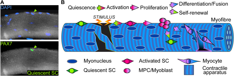
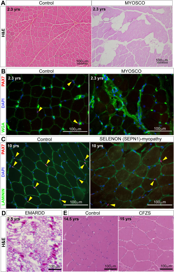
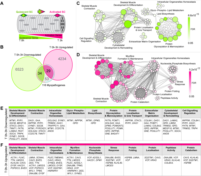
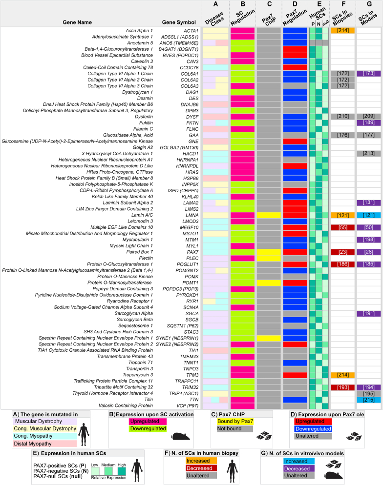
Muscular dystrophies and congenital myopathies arise from specific genetic mutations causing skeletal muscle weakness that reduces quality of life. Muscle health relies on resident muscle stem cells called satellite cells, which enable life-course muscle growth, maintenance, repair and regeneration. Such tuned plasticity gradually diminishes in muscle diseases, suggesting compromised satellite cell function. A central issue however, is whether the pathogenic mutation perturbs satellite cell function directly and/or indirectly via an increasingly hostile microenvironment as disease progresses. Here, we explore the effects on satellite cell function of pathogenic mutations in genes (myopathogenes) that associate with muscle disorders, to evaluate clinical and muscle pathological hallmarks that define dysfunctional satellite cells. We deploy transcriptomic analysis and comparison between muscular dystrophies and myopathies to determine the contribution of satellite cell dysfunction using literature, expression dynamics of myopathogenes and their response to the satellite cell regulator PAX7. Our multimodal approach extends current pathological classifications to define Satellite Cell-opathies: muscle disorders in which satellite cell dysfunction contributes to pathology. Primary Satellite Cell-opathies are conditions where mutations in a myopathogene directly affect satellite cell function, such as in Progressive Congenital Myopathy with Scoliosis (MYOSCO) and Carey-Fineman-Ziter Syndrome (CFZS). Primary satellite cell-opathies are generally characterised as being congenital with general hypotonia, and specific involvement of respiratory, trunk and facial muscles, although serum CK levels are usually within the normal range. Secondary Satellite Cell-opathies have mutations in myopathogenes that affect both satellite cells and muscle fibres. Such classification aids diagnosis and predicting probable disease course, as well as informing on treatment and therapeutic development.

Keywords: Congenital myopathy; Muscle stem cell; Muscular dystrophy; Myopathogene; PAX7; Satellite Cell-opathy; Satellite cell; Skeletal muscle.


Figures

Fig. 1
Fig. 1
Satellite cell myogenesis. A. Representative image of a human quadriceps myofibre showing a quiescent satellite cell expressing PAX7 (green arrowheads; green nucleus, bottom) with myonuclei (blue) stained with DAPI. B. Satellite cells (SC) normally lay mitotically quiescent (green) between the basal lamina and plasmalemma of most myofibres. In response to stimuli (growth, trauma, disease), quiescent satellite cells are activated (red) and proliferate to generate a population of myoblasts (pink - lilac). A proportion of myoblasts undergo self-renewal to replenish the quiescent pool (green) on the myofibre, ensuring future stem cell function. Other myoblasts enter differentiation, becoming myocytes (lilac - blue) that fuse to contribute new myonuclei (blue) to pre-existing multinucleated muscle fibres or fuse together to form new myofibres.
Fig. 2
Fig. 2
Myopathogenes causing muscular dystrophies and myopathies and the subset within the Gene Ontology Satellite Cell term with their associated muscle disorders. A. Distribution of the 116 myopathogenes [15] between human inherited muscular dystrophies (MDs), congenital muscular dystrophies (CMDs), congenital myopathies (CMPs) and distal myopathies (DMs). B. Venn diagram showing the number of myopathogenes shared between the selected muscle disease groupings. C. Gene Ontologies (GOs) containing ‘Satellite Cell’ comprising the term Gene Ontology Satellite Cell (GOSC). D. This novel GOSC contains 30 satellite cell-associated annotated genes. E. Venn diagram showing that four of the myopathogenes involved in satellite cell function in GOSC are associated with muscular dystrophy/myopathy when mutated: PAX7, SELENON (formerly SEPN1), MEGF10 and CAPN3 (green overlap). F. Predominant muscle groups (dotted black line depicts diaphragm) affected by mutations in PAX7 (MYOSCO), SELENON (RSMD1), MEGF10 (EMARDD) and CAPN3 (LGMD1R). Each disorder is colour coded as per A and B to indicate category.
Fig. 3
Fig. 3
Muscle pathology in MYOSCO, SELENON (SEPN1)-related, EMARDD and CFZS. A. Representative Haematoxylin and Eosin (H&E) staining on MYOSCO quadriceps muscle biopsy exhibiting myopathology, notably areas of fat infiltration, compared to age-matched unaffected muscle (Control). B. Representative PAX7 immunolabelling demonstrating absence of satellite cells (PAX7-positive cells are red, indicated by yellow arrowheads) in MYOSCO compared to age-matched unaffected muscle (Control). Myofibres are delimited by Wheat germ agglutinin (WGA, green) and nuclei stained with DAPI (blue). C. Representative PAX7 immunolabelling showing reduction of satellite cells (PAX7-positive cells are red, indicated by yellow arrowheads) in SELENON (SEPN1)-myopathy compared to age-matched unaffected muscle (Control). Myofibres delimited by LAMININ (green) and nuclei identified with DAPI (blue). D. Representative H&E staining on an EMARDD muscle biopsy illustrating variation in myofibre size and fat/fibrotic infiltration. E. Representative H&E staining on CFZS muscle highlighting myofibre hypertrophy compared to age-matched unaffected muscle (Control) (adapted from Ref. [98]). The age of subjects at the time of muscle biopsy are reported, together with 100 μm scale bars.
Fig. 4
Fig. 4
Ontological analysis of the 63 myopathogenes differentially expressed during early satellite cell activation. A. Schematic of quiescent/activated satellite cell gene-sets [168] used to explore involvement of the 116 myopathogenes in satellite cell biology. Diagram shows transcriptomic changes occurring during early murine satellite cell activation, where genes involved in quiescence (green, T 0h) are likely to be downregulated in parallel with upregulation of genes supporting satellite cell activation (magenta, T 3h). B. Venn diagram illustrating overlap between significant differentially expressed genes (downregulated, green and upregulated, magenta) during the first 3 hours (T 0h vs T 3h) of satellite cell activation (A) and the 116 myopathogenes [15]. C and D. Bubble plots demonstrating networks of enriched Gene Ontologies (GOs), clustered according to main biological processes, of differentially expressed myopathogenes during early satellite cell dynamics. C. Main biological processes enriched in downregulated myopathogenes (green). D. Main biological processes enriched in upregulated myopathogenes (magenta). GOs (bubbles) were retrieved using Metascape (metascape.org; [240]) and Panther Gene Ontology (geneontology.org; [241]) and visualised using Cytoscape (cytoscape.org; v3.8.2; [242]). Layout parameters were optimised for presentation. Bubbles are coloured based on False Discovery Rate (FDR) values and size is proportional to number of genes within specific GO terms, grey lines represent genes shared across different GOs. E and F. Differentially expressed (down, green E; up, magenta F) satellite cell-myopathogenes annotated within main biological process (GOs clusters).
Fig. 5
Fig. 5
Summary of myopathogenes associated with satellite cell function. Heatmap depicting the characteristics of the 63 myopathogenes associated with satellite cells. A. Mutation in specific neuromuscular disease class/es [15]. B. Differential expression during early satellite cell (SC) activation in mouse [168]. C. Binding by Pax7 in chromatin immunoprecipitation (ChIP) in mouse myoblasts [169]. D. Differential regulation in response to Pax7 overexpression (o/e) in mouse myoblasts ([169]; LogFold Change≥ 0.5 or ≤ −0.5). E. Expression in PAX7-positive, PAX7-negative or PAX7-null human satellite cells ([24] reported as low, medium or high, based on counts per million (cpm). F. Effect of the myopathogene mutation on the number of satellite cells in patients with citation. G. Effect of the myopathogene mutation on satellite cells in in vivo/vitro models with citation. Sample type and species source of data from human, mouse or cultured cells are depicted by icons in the key.

References

    1. Janssen I., Heymsfield S.B., Wang Z.M., Ross R. Skeletal muscle mass and distribution in 468 men and women aged 18-88 yr. J. Appl. Physiol. 2000;89:81–88. 1985. - PubMed
    1. Zammit P.S. Function of the myogenic regulatory factors Myf5, MyoD, Myogenin and MRF4 in skeletal muscle, satellite cells and regenerative myogenesis. Semin. Cell Dev. Biol. 2017;72:19–32. - PubMed
    1. Chal J., Pourquie O. Making muscle: skeletal myogenesis in vivo and in vitro. Development. 2017;144:2104–2122. - PubMed
    1. Relaix F., Bencze M., Borok M.J., Der Vartanian A., Gattazzo F., Mademtzoglou D., Perez-Diaz S., Prola A., Reyes-Fernandez P.C., Rotini A., Taglietti t. Perspectives on skeletal muscle stem cells. Nat. Commun. 2021;12:692. - PMC - PubMed
    1. Attwaters M., Hughes S.M. Cellular and molecular pathways controlling muscle size in response to exercise. FEBS J. 2021 doi: 10.1111/febs.15820. Online ahead of print. - DOI - PubMed

Publication types

MeSH terms