

Correlation of 68Ga-FAPi-46 PET Biodistribution with FAP Expression by Immunohistochemistry in Patients with Solid Cancers: Interim Analysis of a Prospective Translational Exploratory Study
- PMID: 34740953
- PMCID: PMC9258565
- DOI: 10.2967/jnumed.121.262426
Correlation of 68Ga-FAPi-46 PET Biodistribution with FAP Expression by Immunohistochemistry in Patients with Solid Cancers: Interim Analysis of a Prospective Translational Exploratory Study
Abstract
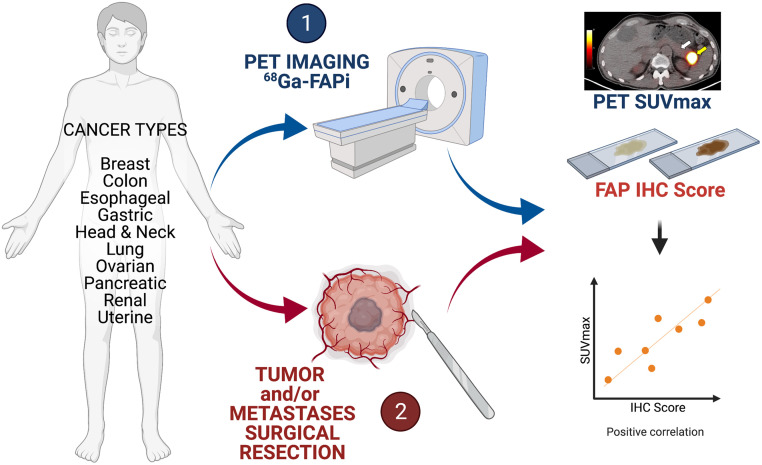
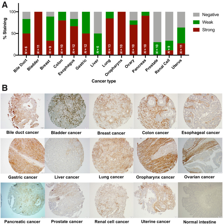
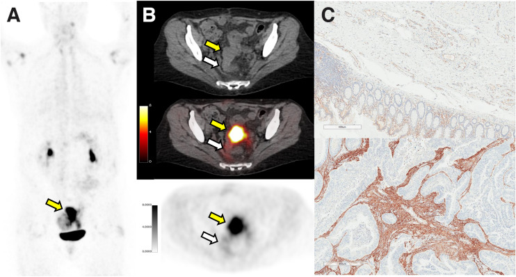
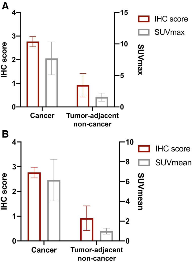
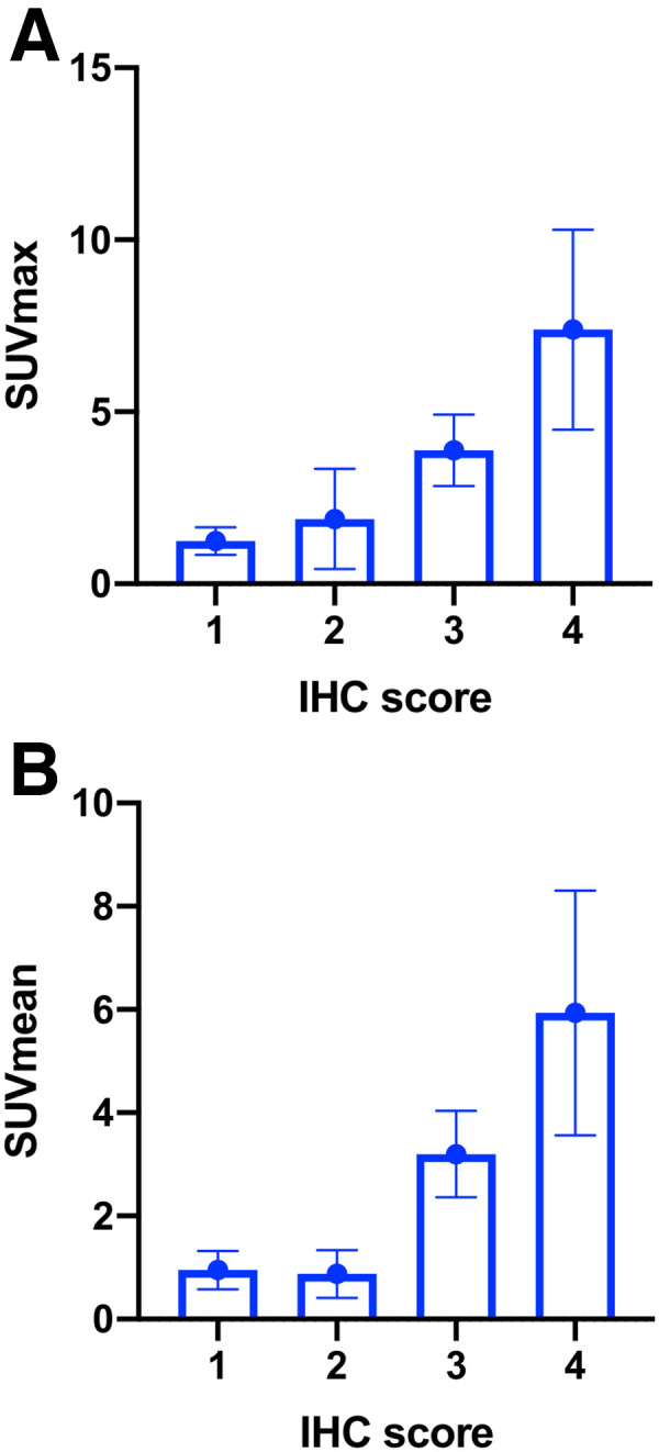
Fibroblast activation protein (FAP)-expressing cancer-associated fibroblasts confer treatment resistance and promote metastasis and immunosuppression. Because FAP is overexpressed in many cancers, radiolabeled molecules targeting FAP are studied for their use as pancancer theranostic agents. This study aimed to establish the spectrum of FAP expression across various cancers by immunohistochemistry and to explore whether 68Ga FAP inhibitor (FAPi)-46 PET biodistribution faithfully reflects FAP expression from resected cancer and non-cancer specimens. Methods: We conducted a FAP expression screening using immunohistochemistry on a pancancer human tissue microarray (141 patients, 14 different types of cancer) and an interim analysis of a prospective exploratory imaging trial in cancer patients. Volunteer patients underwent 1 whole-body 68Ga-FAPi-46 PET/CT scan and, subsequently, surgical resection of their primary tumor or metastasis. 68Ga-FAPi-46 PET SUVmax and SUVmean was correlated with FAP immunohistochemistry score in cancer and tumor-adjacent non-cancer tissues for each patient. Results: FAP was expressed across all 14 cancer types on tissue microarray with variable intensity and frequency, ranging from 25% to 100% (mean, 76.6% ± 25.3%). Strong FAP expression was observed in 50%-100% of cancers of the bile duct, bladder, colon, esophagus, stomach, lung, oropharynx, ovary, and pancreas. Fifteen patients with various cancer types (colorectal [n = 4], head and neck [n = 3], pancreas [n = 2], breast [n = 2], stomach [n = 1], esophagus [n = 2], and uterus [n = 1]) underwent surgery after their 68Ga-FAPi-46 PET/CT scan within a mean interval of 16.1 ± 14.4 d. 68Ga-FAPi-46 SUVs and immunohistochemistry scores were higher in cancer than in tumor-adjacent non-cancer tissue: mean SUVmax 7.7 versus 1.6 (P < 0.001), mean SUVmean 6.2 versus 1.0 (P < 0.001), and mean FAP immunohistochemistry score 2.8 versus 0.9 (P < 0.001). FAP immunohistochemistry scores strongly correlated with 68Ga-FAPi 46 SUVmax and SUVmean: r = 0.781 (95% CI, 0.376-0.936; P < 0.001) and r = 0.783 (95% CI, 0.379-0.936; P < 0.001), respectively. Conclusion: In this interim analysis of a prospective exploratory imaging trial, 68Ga-FAPi-46 PET biodistribution across multiple cancers strongly correlated with FAP tissue expression. These findings support further exploration of FAPi PET as a pancancer imaging biomarker for FAP expression and as a stratification tool for FAP-targeted therapies.
Keywords: 68Ga-FAPi-46; PET/CT; cancer; fibroblast activation protein; immunohistochemistry.
© 2022 by the Society of Nuclear Medicine and Molecular Imaging.
Figures
References
-
- Fearon DT. The carcinoma-associated fibroblast expressing fibroblast activation protein and escape from immune surveillance. Cancer Immunol Res. 2014;2:187–193. - PubMed
-
- Kraman M, Bambrough PJ, Arnold JN, et al. . Suppression of antitumor immunity by stromal cells expressing fibroblast activation protein-alpha. Science. 2010;330:827–830. - PubMed
Publication types
MeSH terms
Substances
Grants and funding
LinkOut - more resources
Full Text Sources
Other Literature Sources
Medical
Miscellaneous