. 2021 Dec 1;207(11):2785-2798.
doi: 10.4049/jimmunol.2100107. Epub 2021 Nov 5.

β-Glucan Induces Distinct and Protective Innate Immune Memory in Differentiated Macrophages

Affiliations

β-Glucan Induces Distinct and Protective Innate Immune Memory in Differentiated Macrophages

Cody L Stothers et al. J Immunol. .

Abstract

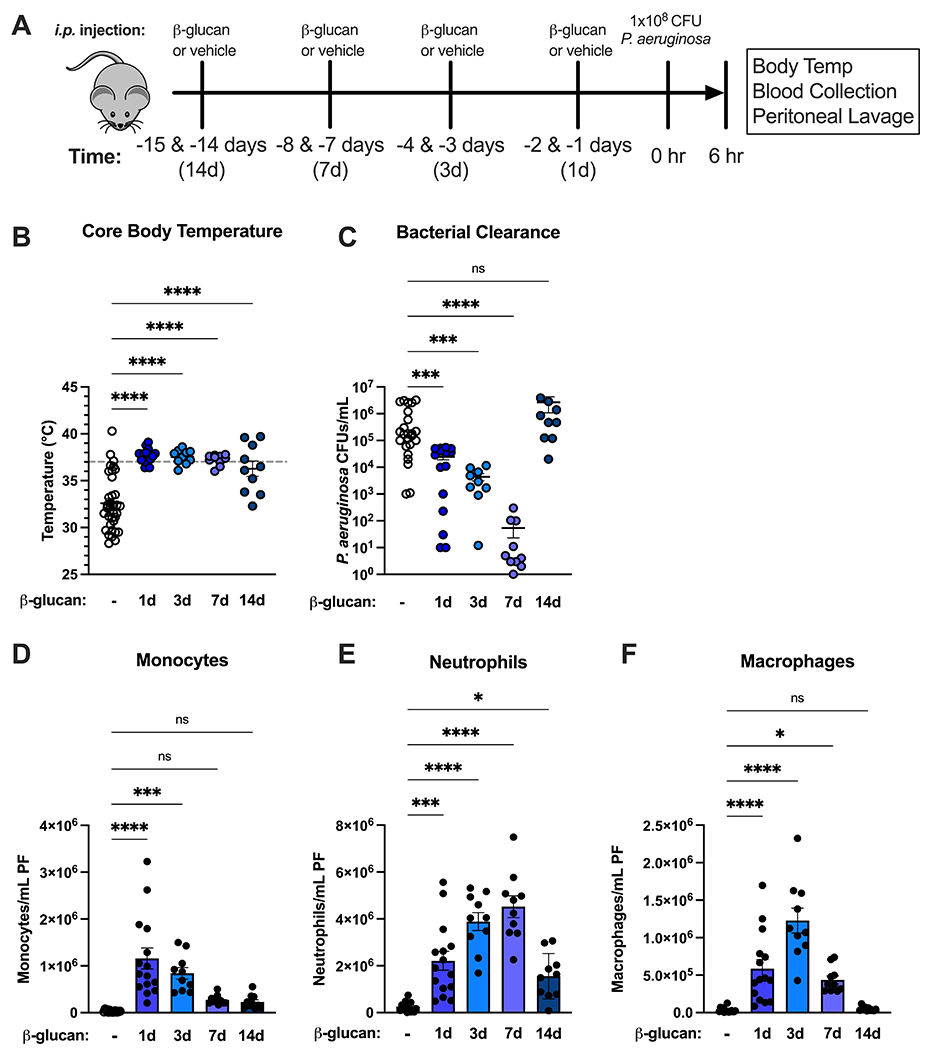
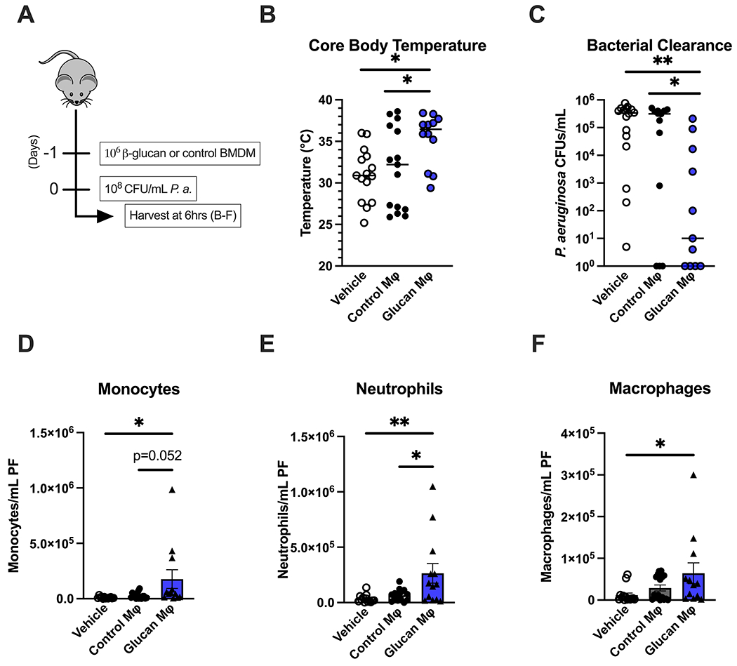
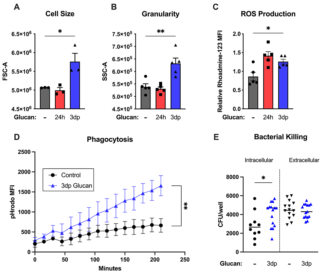
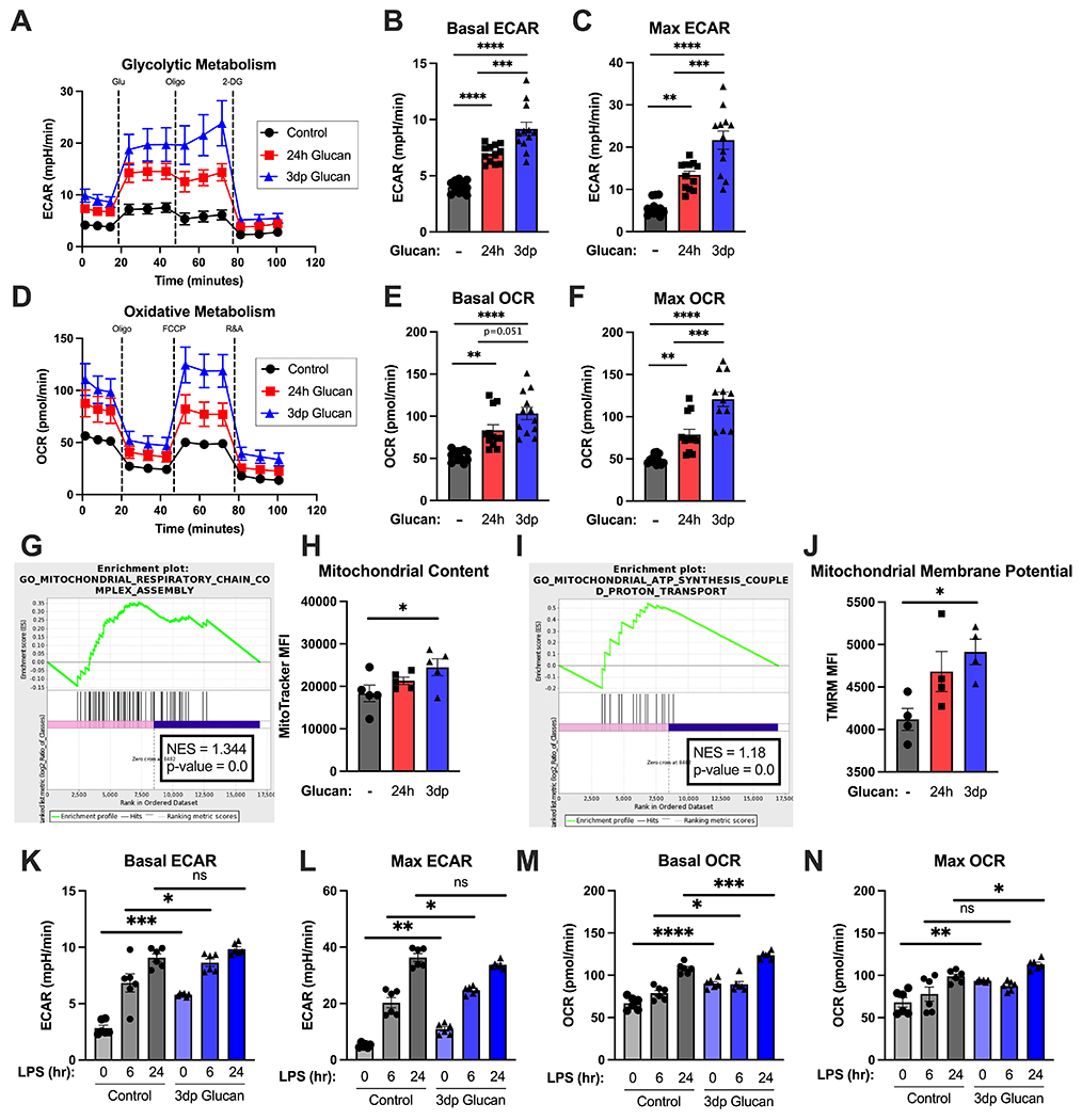
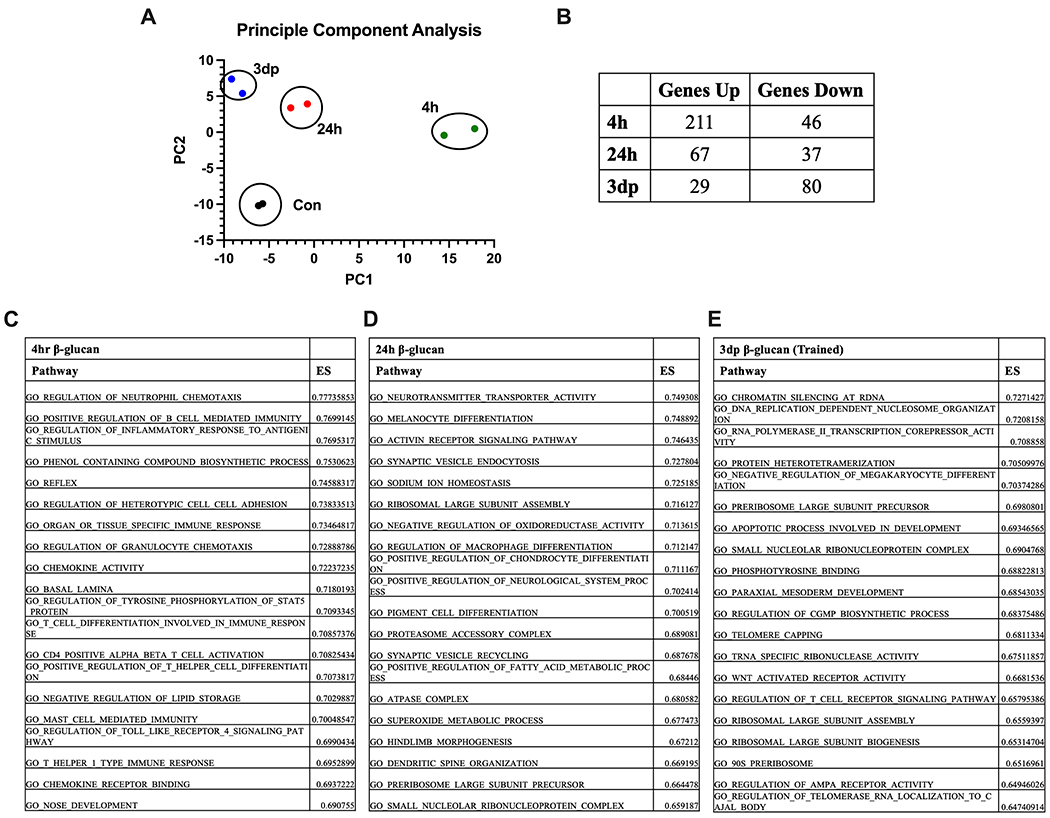
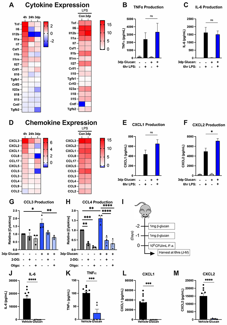
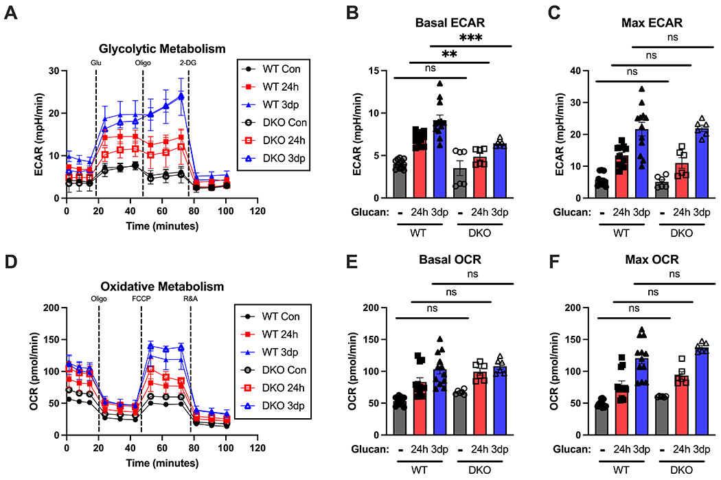
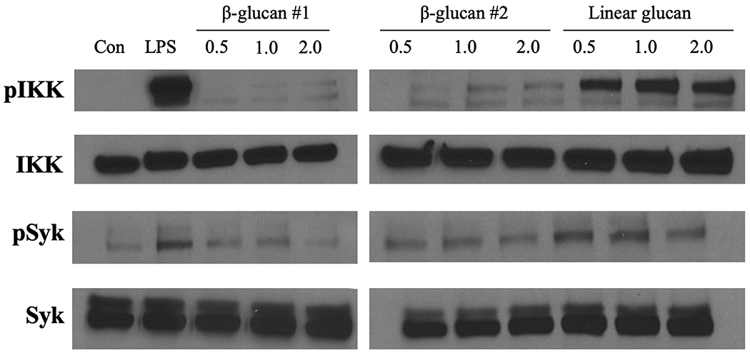
Bacterial infections are a common and deadly threat to vulnerable patients. Alternative strategies to fight infection are needed. β-Glucan, an immunomodulator derived from the fungal cell wall, provokes resistance to infection by inducing trained immunity, a phenomenon that persists for weeks to months. Given the durability of trained immunity, it is unclear which leukocyte populations sustain this effect. Macrophages have a life span that surpasses the duration of trained immunity. Thus, we sought to define the contribution of differentiated macrophages to trained immunity. Our results show that β-glucan protects mice from Pseudomonas aeruginosa infection by augmenting recruitment of innate leukocytes to the site of infection and facilitating local clearance of bacteria, an effect that persists for more than 7 d. Adoptive transfer of macrophages, trained using β-glucan, into naive mice conferred a comparable level of protection. Trained mouse bone marrow-derived macrophages assumed an antimicrobial phenotype characterized by enhanced phagocytosis and reactive oxygen species production in parallel with sustained enhancements in glycolytic and oxidative metabolism, increased mitochondrial mass, and membrane potential. β-Glucan induced broad transcriptomic changes in macrophages consistent with early activation of the inflammatory response, followed by sustained alterations in transcripts associated with metabolism, cellular differentiation, and antimicrobial function. Trained macrophages constitutively secreted CCL chemokines and robustly produced proinflammatory cytokines and chemokines in response to LPS challenge. Induction of the trained phenotype was independent of the classic β-glucan receptors Dectin-1 and TLR-2. These findings provide evidence that β-glucan induces enhanced protection from infection by driving trained immunity in macrophages.


Conflict of interest statement

The authors declare no conflict of interest.

Figures

Figure 1.
Figure 1.. Beta-glucan augments innate immune defense against P. aeruginosa in mice.
(A) C57Bl/6 mice were injected i.p. with β-glucan (1 mg) or vehicle on 2 consecutive days at 1, 3, 7 and 14 days prior to i.p. inoculation with 108 CFU P. aeruginosa with harvest of plasma and peritoneal lavage fluid 6 hours after infection. (B) Core (rectal) body temperature in vehicle- or β-glucan-treated mice. (C) CFUs of P. aeruginosa per mL of peritoneal fluid. (D-F) Number of monocytes (D), neutrophils (E), or macrophages (F) in infected vehicle- or β-glucan-treated mice. Body temperature and clearance data shown as median. All other data shown as mean ± SEM. N = 10-20 mice per group. * p<0.05, ** p<0.01, *** p<0.001, **** p<0.0001 by Mann-Whitney U test (B, C) or ANOVA with Tukey’s post-hoc multiple comparison test (D-F).
Figure 2.
Figure 2.. Adoptive transfer of β-glucan-trained macrophages protects against P. aeruginosa infection.
(A) BMDM were treated with β-glucan (5 μg) or vehicle for 24 hours, washed and allowed to rest for 3 days. C57Bl/6 mice were injected i.p. with vehicle (PBS), control BMDM, or β-glucan-treated BMDM 24 hours prior to i.p. inoculation with 108 CFU P. aeruginosa with subsequent harvest of plasma and peritoneal lavage fluid 6 hours later. (B) Core (rectal) body temperature in vehicle, control-BMDM, or β-glucan-BMDM-treated mice after P. aeruginosa challenge. (C) CFU of P. aeruginosa per mL of peritoneal fluid. (D-F) Number of monocytes (D), neutrophils (E), or macrophages (F) in vehicle, control- or β-glucan-BMDM-treated mice. Body temperature and clearance data shown as median. All other data shown as mean ± SEM. N = 10-15 mice per group. * p<0.05, ** p<0.01 by Kruskal Wallis test with Dunn’s post-hoc multiple comparison test (B, C) or ANOVA with Tukey’s post-hoc multiple comparison test (D-F).
Figure 3.
Figure 3.. Beta-glucan-trained macrophages display a robust antimicrobial phenotype.
BMDM were treated with β-glucan (5 μg) or vehicle for 24 hours (24h), washed and allowed to rest for 3 days (3dp) followed by assessment of macrophage phenotype. (A) Cell size was measured by forward scatter. (B) Cell granularity as measured by side scatter. (C) Rhodamine-123 fluorescence was measured after a 15-minute incubation period using flow cytometry. (D) Control or trained BMDM were incubated with pHrodo S. aureus particles. pHrodo MFI was measured every 15 minutes for 5 hours. (E) BMDM were incubated with P. aeruginosa. Intracellular and extracellular CFU were quantified as described in Methods. Data shown as mean ± SEM. Experiments were performed with 3-5 biological replicates. * p<0.05, ** p<0.01 by ANOVA with Tukey’s post-hoc multiple comparison test (A-C) or repeated two-way ANOVA (D).
Figure 4.
Figure 4.. Beta-glucan training augments metabolism and increases mitochondrial content and membrane potential in macrophages.
BMDM were treated with β-glucan (5 μg) or vehicle for 24 hours (24h), washed and allowed to rest for 3 days (3dp) followed by assessment of macrophage metabolic phenotype. (A) Glycolysis stress test of control, 24h, and trained (3dp) BMDM on the Seahorse XFe96. Extracellular acidification rate was measured over time at baseline and after glucose, oligomycin, and 2-deoxyglucose addition. (B) Basal ECAR as determined from 3 separate runs. (C) Maximum ECAR as determined from 3 separate runs. (D) Oxidative stress test of control, 24h, and trained BMDM on the Seahorse XFe96. Oxygen consumption rate was measured over time at baseline and after oligomycin, FCCP, and R&A administration. (E) Basal OCR as determined from 3 separate runs. (F) Maximum OCR as determined from 3 separate runs. (G) Enrichment plot for GO term “Mitochondrial Respiratory Chain Complex Assembly” for control vs. trained (3dp) BMDM. (H) MitoTracker Green MFI was measured in BMDM by flow cytometry after 30 minutes incubation. (I) Enrichment plot for GO term “Mitochondrial ATP Synthesis-Coupled Proton Transport” for control vs. Trained (3dp) BMDM. (J) TMRM MFI was measured in BMDM by flow cytometry after 30 minutes incubation. (K-L) Basal and maximal ECAR as determined by Seahorse XFe96 in control and trained BMDM stimulated with vehicle or 100 ng/mL LPS for 4hr and 24hr. (M-N) Basal and maximal OCR as determined by Seahorse XFe96 in control and trained BMDM stimulated with vehicle or 100 ng/mL LPS for 4hr and 24hr. Data shown as mean ± SEM. Experiments were performed with 3-5 biological replicates. * p<0.05, ** p<0.01, *** p<0.001, **** p<0.0001 by ANOVA with Tukey’s post-hoc multiple comparison test.
Figure 5.
Figure 5.. Beta-glucan induces a distinct transcriptomic profile in macrophages.
BMBM were treated with β-glucan (5 μg) or vehicle for 4 (4h) or 24 (24h) hours. A subset of 24h BMDM were washed and allowed to rest for 3 days (3dp). Gene expression was measured using RNAseq. (A) Principal Component Analysis of BMDM harvested at specified time points. (B) Relative gene expression by BMDM treated with β-glucan relative to control at the specified time points. ES = enrichment score (C) Top 20 pathways identified by gene ontology analysis at 4 hours after β-glucan treatment relative to control. (D) Top 20 pathways identified by gene ontology analysis at 24 hours after β-glucan treatment relative to control. (E) Top 20 pathways identified by gene ontology analysis at 3dp after β-glucan treatment relative to control. RNA quantification performed in duplicate.
Figure 6.
Figure 6.. Beta-glucan alters macrophage cytokine and chemokine production.
BMBM were treated with β-glucan (5 μg) or vehicle for 4 (4h) or 24 (24h) hours. A subset of 24h BMDM were washed and allowed to rest for 3 days (3dp). Cytokine mRNA expression was measured by RNAseq, protein was measured by ELISA. (A) Heatmaps of cytokine mRNA expression by BMDM at 4h, 24h, or 3dp relative to control and by control and 3dp BMDM at 4h after LPS challenge. (B-C) Concentrations of TNFα and IL-6 in conditioned media from control and 3dp BMDM before and after LPS challenge. (D) Heatmaps of chemokine mRNA expression by BMDM at 4h, 24h, or 3dp relative to control and by control and 3dp BMDM at 4h after LPS challenge. (E-F) Concentrations of CXCL1 and CXCL2 in conditioned media from control and 3dp BMDM before and after LPS challenge. (G-H) Concentrations of CCL3 and CCL4 in conditioned media from control and 3dp BMDM after treatment with 2-DG or oligomycin for 6 hours. Data normalized to untreated control BMDM. (I) C57Bl/6 mice were injected i.p. with β-glucan (1 mg) or vehicle on 2 consecutive days prior to i.p. inoculation with 108 CFU P. aeruginosa with subsequent harvest of plasma 6 hours later. (J-M) Concentrations of IL-6, TNFα, CXCL1 and CXCL2 in peritoneal lavage at 6 hours after P. aeruginosa challenge. RNASeq experiments were performed in duplicate. All other in vitro experiments were performed with 3-5 biological replicates. N=5 mice per group for in vivo experiments. * p<0.05, ** p<0.01, *** p<0.001, **** p<0.0001 by ANOVA with Tukey’s post-hoc multiple comparison test.
Figure 7.
Figure 7.. Beta-glucan-induced protection against P. aeruginosa is independent of Dectin-1 and Toll-like receptor (TLR)-2.
(A) Wildtype, Dectin-1 knockout, TLR-2, or Dectin-1/TLR2 double knockout C57Bl/6 mice were injected i.p. with β-glucan (1 mg) or vehicle 48 and 24 hours prior to i.p. inoculation with 108 CFU P. aeruginosa with subsequent harvest of plasma and peritoneal lavage fluid 6 hours later. (B) Core (rectal) body temperature in vehicle- or β-glucan treated mice 6 hours after i.p. P. aeruginosa. (C) CFUs of P. aeruginosa per mL of peritoneal fluid. (D-F) Number of monocytes (D), neutrophils (E), or macrophages (F) in infected vehicle- or β-glucan-treated mice. Body temperature and clearance data shown with median. All other data shown as mean ± SEM. N = 10-15 mice per group. * p<0.05, ** p<0.01, *** p<0.001, **** p<0.0001 by Kruskal-Wallis test followed by Dunn’s post hoc multiple comparison test (B, C) or ANOVA with Tukey’s post-hoc multiple comparison test (D-F).
Figure 8.
Figure 8.. Metabolic alterations in β-glucan-trained macrophages are independent of Dectin-1 and Toll-like receptor 2.
BMDM from WT and DKO mice were treated with β-glucan (5 μg) or vehicle for 24 hours (24h), washed and allowed to rest for 3 days (3dp) followed by assessment of macrophage metabolic phenotype. (A) Glycolysis stress test of control, 24h, and 3dp BMDM on the Seahorse XFe96 in wildtype and DKO mice. Extracellular acidification rate was measured over time at baseline and after glucose, oligomycin, and 2-deoxyglucose administration. (B) Basal ECAR in each group from multiple replicates. (C) Maximum ECAR in each group from multiple replicates. (D) Oxidative stress test of control, 24h, and trained BMDM on the Seahorse Xfe96. Oxygen consumption rate was measured over time at baseline and after oligomycin, FCCP, and R&A administration. (E) Basal OCR in each group from multiple replicates. (F) Maximum OCR in each group from multiple replicates. Data shown as mean ± SEM. Experiments were performed with 3 biological replicates. ** p<0.01, *** p<0.001 by ANOVA with Tukey’s post-hoc multiple comparison test.
Figure 9.
Figure 9.. The β-glucan training reagent induces weak Dectin-1 and Toll-like receptor 2 activation.
Protein was isolated from BMDM treated with β-glucan for 0.5, 1.0, or 2.0 hours. BMDM treated with 100 ng/mL LPS for 1 hour or linear glucan for 0.5, 1.0, or 2.0 hours served as positive controls. Western blot of phosphorylated IkB kinase (pIkK), total IkK (IkK), phosphorylated spleen tyrosine kinase (pSyk), and total Syk (Syk). Blots are representative of three repeated experiments.

References

    1. Mayr FB, Yende S, and Angus DC. 2014. Epidemiology of severe sepsis. Virulence 5: 4–11. - PMC - PubMed
    1. Vincent JL, Rello J, Marshall J, Silva E, Anzueto A, Martin CD, Moreno R, Lipman J, Gomersall C, Sakr Y, Reinhart K, and E. I. G. o. Investigators. 2009. International study of the prevalence and outcomes of infection in intensive care units. JAMA 302: 2323–2329. - PubMed
    1. Manners C, Larios Bautista E, Sidoti H, and Lopez OJ. 2020. Protective Adaptive Immunity Against Severe Acute Respiratory Syndrome Coronaviruses 2 (SARS-CoV-2) and Implications for Vaccines. Cureus 12: e8399. - PMC - PubMed
    1. Nogales A, and DeDiego ML. 2020. Influenza Virus and Vaccination. Pathogens 9. - PMC - PubMed
    1. Gourbal B, Pinaud S, Beckers GJM, Van Der Meer JWM, Conrath U, and Netea MG. 2018. Innate immune memory: An evolutionary perspective. Immunol Rev 283: 21–40. - PubMed

Publication types