Review
. 2022 Jul 13;43(4):678-719.
doi: 10.1210/endrev/bnab039.

Comprehensive Review of Uterine Fibroids: Developmental Origin, Pathogenesis, and Treatment

Affiliations
Review

Comprehensive Review of Uterine Fibroids: Developmental Origin, Pathogenesis, and Treatment

Qiwei Yang et al. Endocr Rev. .

Erratum in

Abstract

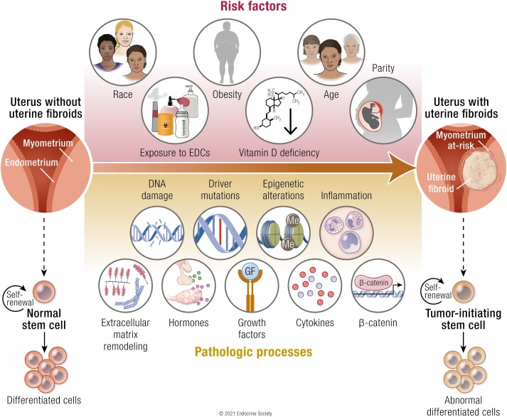
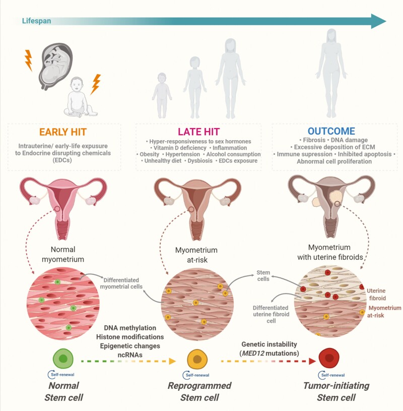
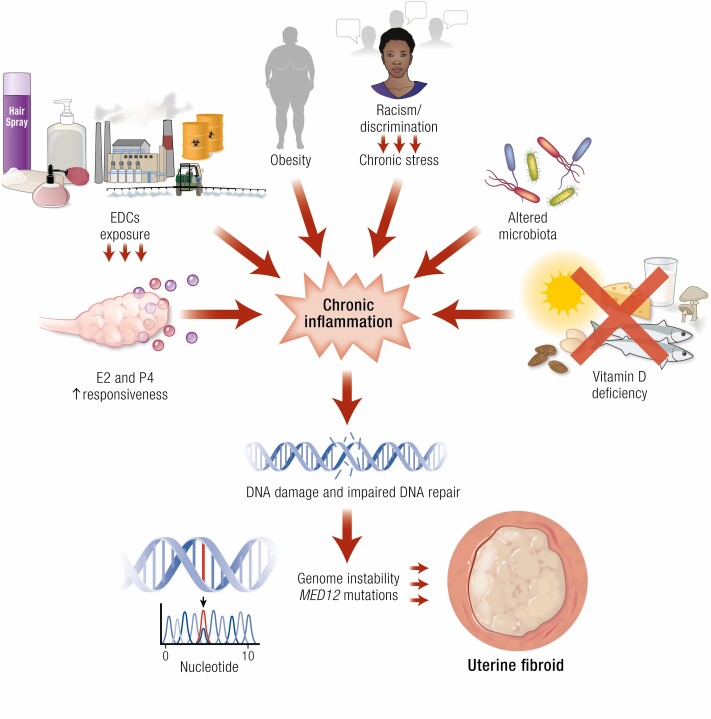
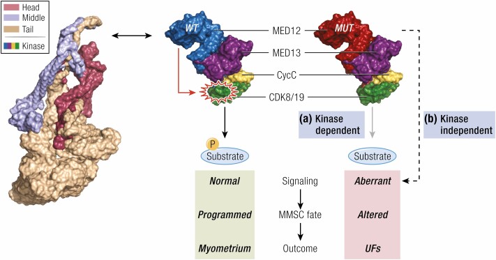
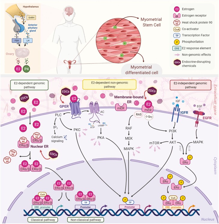
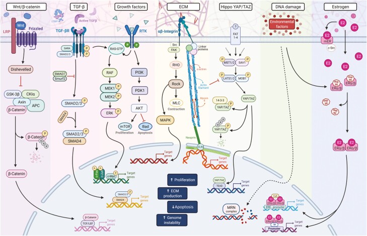
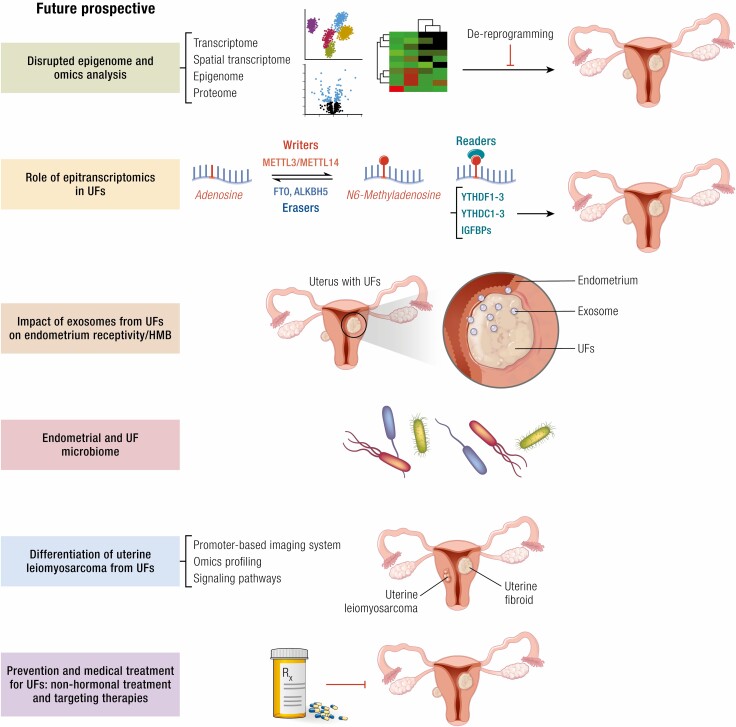
Uterine fibroids are benign monoclonal neoplasms of the myometrium, representing the most common tumors in women worldwide. To date, no long-term or noninvasive treatment option exists for hormone-dependent uterine fibroids, due to the limited knowledge about the molecular mechanisms underlying the initiation and development of uterine fibroids. This paper comprehensively summarizes the recent research advances on uterine fibroids, focusing on risk factors, development origin, pathogenetic mechanisms, and treatment options. Additionally, we describe the current treatment interventions for uterine fibroids. Finally, future perspectives on uterine fibroids studies are summarized. Deeper mechanistic insights into tumor etiology and the complexity of uterine fibroids can contribute to the progress of newer targeted therapies.

Keywords: developmental origin; epigenetics pathways; future directions; genetic instability; novel treatment; reprogramming; uterine fibroids.


Figures

Graphical Abstract
Graphical Abstract
Figure 1.
Figure 1.
Developmental origin of fibroids from myometrial stem cells. Intrauterine and early-life adverse environmental exposure to endocrine-disrupting chemicals may act as the early hit to induce normal myometrial stem cells’ reprogramming by hijacking epigenomic plasticity. The plasticity of the developing epigenome is susceptible to epigenomic changes in myometrial stem cells following later-life adverse exposures, thereby leading to mutations and their transformation into tumor-initiating stem cells. The development and growth of fibroids are mainly characterized by abnormal cell proliferation, inhibited apoptosis, DNA instability, excessive deposition of ECM, and other critical biological pathways. Abbreviations: ECM, extracellular matrix; MED12, RNA polymerase II transcriptional mediator complex subunit 12; ncRNAs, non-coding RNA.
Figure 2.
Figure 2.
Risk factors for uterine fibroids that mainly affect inflammation, DNA damage pathways, and genetic instability. External and internal factors, such as EDC exposure, hyper-responsiveness to sex steroid hormones, obesity, vitamin D deficiency, and altered reproductive tract microbiome, contribute to chronic systemic inflammation. The inflammatory environment, EDC exposure, and vitamin D deficiency promote DNA damage and the accumulation of mutations. Consequently, these genetic events may activate the pathways involved in cell proliferation, the inhibition of apoptosis, and ECM remodeling, ultimately leading to the development and growth of fibroids. Abbreviations: E2, estrogen; EDCs, endocrine-disrupting chemicals; MED12, RNA polymerase II transcriptional mediator complex subunit 12; P4, progesterone.
Figure 3.
Figure 3.
Role of MED12 mutation in the pathogenesis of fibroids. Two mutually compatible models are demonstrating that fibroids driver mutations in MED12 trigger myometrial stem cell transformation and fibroids formation through altered signaling. In the first model (A), MED12 mutations in exon 2 disrupt the CDK8 T-loop conformation to affect Mediator kinase activity and the phosphorylation of downstream targets, including those that control myometrial stem cell fate and/or function. In the second model (B), MED12 mutations alter gene expression programs that control myometrial stem cell fate and/or function through kinase-independent mechanisms, such as MED12 interactions with transcriptional regulatory proteins (173). The 2 models are not mutually exclusive, and both scenarios could contribute to fibroids pathogenesis. Shown here is the 4-subunit Mediator kinase module comprising MED13, MED12, CycC, and CDK8/19 that variably associates with a core Mediator, which is collectively composed of 26 different subunits arranged into 3 structurally defined domains, ie, Head, Middle, and Tail. The structure of the core Mediator is from Clark et al (137), whereas that of the kinase module is from Li et al (167). Abbreviations: CDK8/19, cyclin-dependent kinase 8/19; CycC, cyclin C; MED12/13, RNA polymerase II transcriptional mediator complex subunit 12/13; MMSC, myometrial stem cell; UFs, uterine fibroids.
Figure 4.
Figure 4.
Estrogen receptor-mediated signaling pathways in the myometrium. The biosynthesis of natural E2 occurs in the ovary downstream the actions of the LH and the FSH, which are regulated by the GnRH. E2 mediates its biological response through several pathways, which can be classified as genomic and nongenomic. There are 3 main mechanisms of genomic regulation mediated by ER. Firstly, in the classical pathway, E2 ligands passively enter the cells by diffusion. ERα and ERβ are localized in the cytosol and are attached to the chaperon Hsp90, which is released after binding with estrogen. The estrogen-bound receptors form dimers that enter the nucleus and bind to the ERE, specific DNA sequences of the promoters of target genes affecting their transcription. Secondly, the nonclassical pathway involves binding the E2-bound ER to TFs that are already bound to the DNA. The third mechanism is hormone-independent. The ER can regulate E2 responses by activating the signaling of growth factors via the phosphorylation of different serine (118/167) residues on the receptor. In addition to upregulating gene expression, E2 exerts its nongenomic rapid biological actions by interaction with membrane receptors. GPER, a membrane-integrated 7-transmembrane receptor, activates heterotrimeric G-proteins after binding with estrogen to elicit various nongenomic responses, such as calcium signaling, PKC, and cAMP/PKA pathways. Bound-membrane ERs (ERα, ERβ, ER36, and ER46) also activate cytosolic signalings, such as PI3K/Akt and MAPK. In addition, the activation of kinases results in the phosphorylation of specific transcription factors that regulate gene expression. EDCs are exogenous, manufactured chemicals, such as genistein, bisphenol A, and phthalates that mimic natural estrogen molecular and cellular responses, thereby altering the functions of the endocrine system. These chemicals are associated with the developmental origin of fibroids and their pathogenesis. Abbreviations: AC, adenylyl cyclase; AKT, protein kinase B; cAMP, cyclic AMP; CoA, coactivator; E2, estrogen; EDCs, endocrine-disrupting chemicals; EGFR, epidermal growth factor receptor; ER, estrogen receptor; ERE, estrogen-responsive elements; FSH, follicle-stimulating hormone; GnRH, gonadotropin-releasing hormone; GPER, G-protein coupled estrogen receptor 1; Hsp90, heat shock protein 90; IGFR, insulin-like growth factor 1 receptor; IP3, inositol trisphosphate; LH, luteinizing hormone; MAPK, mitogen-activated protein kinase; MEK, mitogen-activated protein kinase kinase; mER, membrane-bound estrogen receptor; mTOR, mammalian target of rapamycin; PI3K, phosphoinositol-3-kinase; PKA, protein kinase A; PKC, protein kinase; PLC, phospholipase C; Raf, Rapidly Accelerated Fibrosarcoma Kinase; Ras, Ras GTPase; TFs, transcription factors.
Figure 5.
Figure 5.
Critical pathways in uterine fibroids pathogenesis. The WNT/β-catenin, TGF-β, growth factor–regulated signaling, ECM, estrogen signaling, YAP/TAZ, Rho/ROCK, and DNA damage repair pathways play essential roles in fibroids formation and development. In addition, the crosstalk and interaction among these pathways may initiate and trigger uterine fibroids pathogenesis. Abbreviations: AKT, protein kinase B; APC, adenomatous polyposis coli; Bad, BCL2 associated agonist of cell death; CK1α, casein kinase 1 alpha; E2, Estrogen; ECM, extracellular matrix; ERE, estrogen-responsive elements; ERK, extracellular-signal-regulated kinase; ERα/β, estrogen receptor alpha/beta; FAK, focal adhesion kinase; GSK-3β, glycogen synthase kinase 3 beta; Hsp90, heat shock protein 90; LRP, lipoprotein receptor-related protein; MAPK, mitogen-activated protein kinase; MEK, mitogen-activated protein kinase kinase; mER, membrane-bound estrogen; MLC, Myosin regulatory light chain 2; MRN, Mre11-Rad50-Nbs1 complex; mTOR, mechanistic target of rapamycin; P, phosphorylated site; PDK1, 3-phosphoinositide-dependent protein kinase 1; PI3K, phosphatidylinositol 3-kinase; RAF, Rapidly Accelerated Fibrosarcoma kinase; RHO, Ras-homologous; RTK, receptor tyrosine kinases; SMAD, mothers against DPP (decapentaplegic); Src, proto-oncogene tyrosine-protein kinase; TF, transcription factor; TGFβ, transforming growth factor beta; TGFβR, transforming growth factor beta receptor; Wnt, Wingless-related integration site; YAP, Yes-associated protein; TAZ, transcriptional coactivator with PDZ-binding domain.
Figure 6.
Figure 6.
Future research prospects. Studies elucidating the interplay among genes, epigenome, and epitranscriptome in the context of stem cell biology, microbiome, and the interaction between the endometrium and uterine fibroids can advance the knowledge on the pathogenesis of uterine fibroids and are expected to contribute to developing novel therapeutic approaches for the treatment of patients with uterine fibroids. Abbreviations: ALKBH5, ALKB homolog 5; FTO, fat mass and obesity-associated protein; HMB: heavy menstrual bleeding; IGFBPs, insulin-like growth factor binding protein-3; METTL3,14, methyltransferase like 3 and 14; UFs, uterine fibroids; YTHDF1/YTHDC1, YTH domain-containing protein.

References

    1. Al-Hendy A, Myers ER, Stewart E. Uterine fibroids: burden and unmet medical need. Semin Reprod Med. 2017;35(6):473-480. - PMC - PubMed
    1. Wise LA, Laughlin-Tommaso SK. Epidemiology of uterine fibroids: from menarche to menopause. Clin Obstet Gynecol. 2016;59(1):2-24. - PMC - PubMed
    1. Baird DD, Dunson DB, Hill MC, Cousins D, Schectman JM. High cumulative incidence of uterine leiomyoma in black and white women: ultrasound evidence. Am J Obstet Gynecol. 2003;188(1):100-107. - PubMed
    1. Jayes FL, Liu B, Feng L, Aviles-Espinoza N, Leikin S, Leppert PC. Evidence of biomechanical and collagen heterogeneity in uterine fibroids. Plos One. 2019;14(4):e0215646. - PMC - PubMed
    1. Stewart EA, Laughlin-Tommaso SK, Catherino WH, Lalitkumar S, Gupta D, Vollenhoven B. Uterine fibroids. Nat Rev Dis Primers. 2016;2:16043. - PubMed

Publication types