

The semantics of microglia activation: neuroinflammation, homeostasis, and stress
- PMID: 34742308
- PMCID: PMC8571840
- DOI: 10.1186/s12974-021-02309-6
The semantics of microglia activation: neuroinflammation, homeostasis, and stress
Abstract
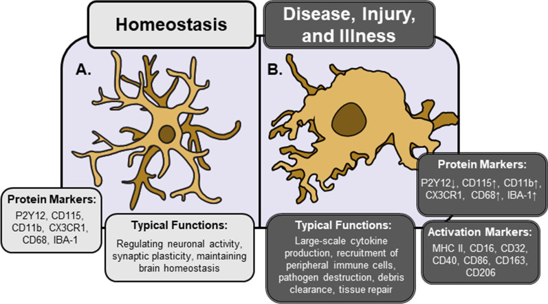
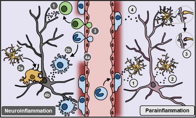
Microglia are emerging as critical regulators of neuronal function and behavior in nearly every area of neuroscience. Initial reports focused on classical immune functions of microglia in pathological contexts, however, immunological concepts from these studies have been applied to describe neuro-immune interactions in the absence of disease, injury, or infection. Indeed, terms such as 'microglia activation' or 'neuroinflammation' are used ubiquitously to describe changes in neuro-immune function in disparate contexts; particularly in stress research, where these terms prompt undue comparisons to pathological conditions. This creates a barrier for investigators new to neuro-immunology and ultimately hinders our understanding of stress effects on microglia. As more studies seek to understand the role of microglia in neurobiology and behavior, it is increasingly important to develop standard methods to study and define microglial phenotype and function. In this review, we summarize primary research on the role of microglia in pathological and physiological contexts. Further, we propose a framework to better describe changes in microglia1 phenotype and function in chronic stress. This approach will enable more precise characterization of microglia in different contexts, which should facilitate development of microglia-directed therapeutics in psychiatric and neurological disease.
Keywords: Depression; Homeostasis; Inflammation; Microglia; Parainflammation; Stress.
© 2021. The Author(s).
Conflict of interest statement
The authors declare no competing interests.
Figures
References
-
- del Río-Hortega BJ. Pío del Río-Hortega: the revolution of Glia. Anat Rec. 2020;303:1232–1241. - PubMed
-
- Murabe Y, Sano Y. Morphological studies on neuroglia—VII. Distribution of “brain macrophages” in brains of neonatal and adult rats, as determined by means of immunohistochemistry. Cell Tissue Res. 1983;229:85–95. - PubMed
-
- Murabe Y, Sano Y. Morphological studies on neuroglia—VI. Postnatal development of microglial cells. Cell Tissue Res. 1982;225:469–485. - PubMed
Publication types
MeSH terms
Grants and funding
- R21-MH120614/foundation for the national institutes of health
- R21 MH120614/MH/NIMH NIH HHS/United States
- R01 MH123545/MH/NIMH NIH HHS/United States
- University of Cincinnati Neurobiology Research Center Pilot Award/college of medicine, university of cincinnati
- 25488/brain and behavior research foundation
LinkOut - more resources
Full Text Sources
Other Literature Sources
Research Materials
