. 2021 Oct 22:12:737616.
doi: 10.3389/fmicb.2021.737616. eCollection 2021.

Seed-Transmitted Bacteria and Fungi Dominate Juvenile Plant Microbiomes

Affiliations

Seed-Transmitted Bacteria and Fungi Dominate Juvenile Plant Microbiomes

David Johnston-Monje et al. Front Microbiol. .

Abstract

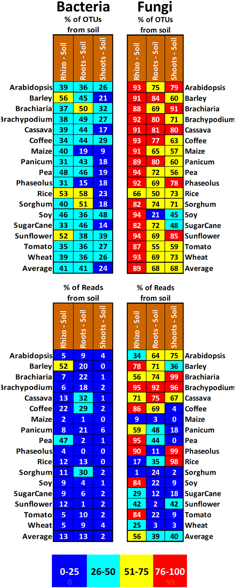
Plant microbiomes play an important role in agricultural productivity, but there is still much to learn about their provenance, diversity, and organization. In order to study the role of vertical transmission in establishing the bacterial and fungal populations of juvenile plants, we used high-throughput sequencing to survey the microbiomes of seeds, spermospheres, rhizospheres, roots, and shoots of the monocot crops maize (B73), rice (Nipponbare), switchgrass (Alamo), Brachiaria decumbens, wheat, sugarcane, barley, and sorghum; the dicot crops tomato (Heinz 1706), coffee (Geisha), common bean (G19833), cassava, soybean, pea, and sunflower; and the model plants Arabidopsis thaliana (Columbia-0) and Brachypodium distachyon (Bd21). Unsterilized seeds were planted in either sterile sand or farm soil inside hermetically sealed jars, and after as much as 60 days of growth, DNA was extracted to allow for amplicon sequence-based profiling of the bacterial and fungal populations that developed. Seeds of most plants were dominated by Proteobacteria and Ascomycetes, with all containing operational taxonomic units (OTUs) belonging to Pantoea and Enterobacter. All spermospheres also contained DNA belonging to Pseudomonas, Bacillus, and Fusarium. Despite having only seeds as a source of inoculum, all plants grown on sterile sand in sealed jars nevertheless developed rhizospheres, endospheres, and phyllospheres dominated by shared Proteobacteria and diverse fungi. Compared to sterile sand-grown seedlings, growth on soil added new microbial diversity to the plant, especially to rhizospheres; however, all 63 seed-transmitted bacterial OTUs were still present, and the most abundant bacteria (Pantoea, Enterobacter, Pseudomonas, Klebsiella, and Massilia) were the same dominant seed-transmitted microbes observed in sterile sand-grown plants. While most plant mycobiome diversity was observed to come from soil, judging by read abundance, the dominant fungi (Fusarium and Alternaria) were also vertically transmitted. Seed-transmitted fungi and bacteria appear to make up the majority of juvenile crop plant microbial populations by abundance, and based on occupancy, there seems to be a pan-angiosperm seed-transmitted core bacterial microbiome. Further study of these seed-transmitted microbes will be important to understand their role in plant growth and health, as well as their fate during the plant life cycle and may lead to innovations for agricultural inoculant development.

Keywords: core microbiome; endophyte; phyllosphere; plant microbiome; plant mycobiome; rhizosphere; seed microbiome; spermosphere.


Conflict of interest statement

The authors declare that the research was conducted in the absence of any commercial or financial relationships that could be construed as a potential conflict of interest.

Figures

FIGURE 1
FIGURE 1
Soil, seeds, and examples of the gnotobiotic terrariums used to grow plants in this study. (A) Fallow cassava field at CIAT where soil was harvested. (B) Cassava field soil after sieving. (C) Soaking in sterile water prior to extraction of seed endosphere and spermosphere DNA (yucca = cassava; maracuya and apple were later replaced with pea and barley). (D) Coffee at harvest after growth in sterile sand on the left and field soil mixed with sand on the right. (E) Barley at harvest after growth in sterile sand on the left and field soil mixed with sand on the right.
FIGURE 2
FIGURE 2
Phylum level classification of bacterial 16S rDNA (A) and fungal ITS DNA (B) amplified from seeds (pies) and spermospheres (donuts) of the 17 different plant species used in this study. OTUs in each repetition were grouped by sample type and color classified by phylum to yield relative proportion by sample. Seeds and spermospheres were added together and sorted by agglomerative hierarchical clustering using Bray–Curtis dissimilarity of phylum distribution.
FIGURE 3
FIGURE 3
Common bacterial 16S and fungal ITS OTUs of (A) seeds and (B) spermospheres. Common OTUs were defined as having an occupancy of more than 60% across seeds or spermospheres of the 17 different plant species. Bacterial OTUs are shown in the top blocks, fungal OTUs on the bottom blocks. Next to each OTU ID# is the predicted genus of that sequence. OTU read proportion is represented by color as shown in the legend, with red squares also showing the proportion as a number.
FIGURE 4
FIGURE 4
Heatmaps of the 40 most common bacterial 16S rDNA and fungal ITS OTUs derived from Miseq analysis of rhizospheres, roots and shoots from 17 different plant species grown in sealed jars on either sterile sand or field soil. Reads were rarified for each sample (fungi—1000/roots and shoots, 2000/rhizospheres; bacteria—3000/roots, 3500/shoots, 6500/rhizospheres), averaged across repetitions, transformed into relative percentages, then clustered by Bray–Curtis dissimilarity. Samples from sand grown plants are labelled in white, samples from soil grown plants are labelled in brown. Cells are shaded by percentage value with 0% being dark blue, up to 0.1% being light blue, between 0.1–0.25% being green, 0.25–0.5% being light yellow, 0.5-1% being dark yellow, 1-5% being orange and greater than 5% being red.
FIGURE 5
FIGURE 5
Proportion of OTUs and reads in the shoots, roots, and rhizospheres of different plant species growing either on sterile sand (labeled in white) or field soil (labeled in brown) that were also observed in that species’ corresponding seeds or spermospheres. Cells are shaded to reflect proportion, with 0–25% being blue, 26–50% being green, 51–75% being yellow, and 76–100% being red. An average across all the plant species is shown at the bottom of each column.
FIGURE 6
FIGURE 6
Proportion of OTUs and reads in the shoots, roots, and rhizospheres of different plant species growing on field soil (labeled in brown) that were not observed in that species’ corresponding shoots, roots, and rhizospheres from plants grown on sterile sand. Cells are shaded to reflect proportion, with 0–25% being blue, 26–50% being green, 51–75% being yellow, and 76–100% being red. An average across all the plant species is shown at the bottom of each column.
FIGURE 7
FIGURE 7
Alpha diversity and phylum-level classification of total (A,C) and common (B,D) OTUs of fungal ITS DNA and bacterial 16S rDNA amplified from seeds, spermospheres, rhizospheres, roots, and shoots. Alpha diversity was measured using Shannon H index and is reported on top of each bar. The proportion of each sample type was calculated by averaging all the samples in each respective group (e.g., average of relative proportions from all seed OTUs). (B,D) Total common OTUs were those that were observed in more than 60% of the shoots/roots/rhizospheres of soil-grown plants, while common soil OTUs were observed in more than 60% of soil-grown samples but not in sterile sand-grown samples. Common seed OTUs were those that were observed in both >60% of soil-grown tissues and in >60% of sterile sand-grown tissues. OTUs are classified at the phylum level and shaded according to the legends at the right.

References

    1. Abdelfattah A., Wisniewski M., Schena L., Tack A. J. M. (2021). Experimental evidence of microbial inheritance in plants and transmission routes from seed to phyllosphere and root. Environ. Microbiol. 23 2199–2214. 10.1111/1462-2920.15392 - DOI - PubMed
    1. Adams P. D., Kloepper J. W. (2002). Effect of host genotype on indigenous bacterial endophytes of cotton (Gossypium hirsutum L.). Plant Soil 240 181–189. 10.1023/A:1015840224564 - DOI
    1. Afzal I., Shinwari Z. K., Sikandar S., Shahzad S. (2019). Plant beneficial endophytic bacteria: mechanisms, diversity, host range and genetic determinants. Microbiol. Res. 221 36–49. - PubMed
    1. Allard S. M., Ottesen A. R., Brown E. W., Micallef S. A. (2018). Insect exclusion limits variation in bacterial microbiomes of tomato flowers and fruit. J. Appl. Microbiol. 125 1749–1760. 10.1111/jam.14087 - DOI - PubMed
    1. Arif I., Batool M., Schenk P. M. (2020). Plant microbiome engineering: expected benefits for improved crop growth and resilience. Trends Biotechnol. 38 1385–1396. 10.1016/j.tibtech.2020.04.015 - DOI - PubMed

LinkOut - more resources