. 2021 Oct 20:12:743920.
doi: 10.3389/fmicb.2021.743920. eCollection 2021.

A New Protocruzia Species (Ciliophora: Protocruziida) Isolated From the Mariana Trench Area

Affiliations

A New Protocruzia Species (Ciliophora: Protocruziida) Isolated From the Mariana Trench Area

Chen Liang et al. Front Microbiol. .

Abstract

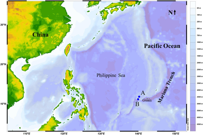
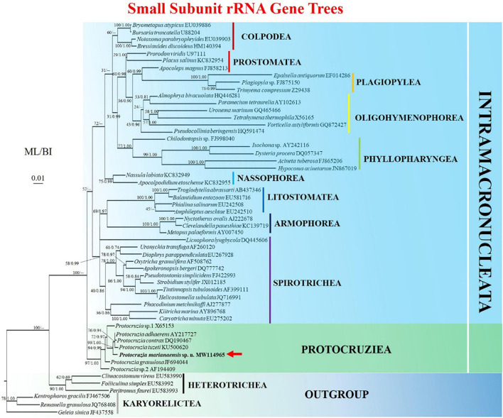
A new species of Protocruzia, isolated from the deep-sea Pacific Ocean (>3,000-m depth) in the vicinity of the Mariana Trench, is described based on morphological and molecular data. The systematic status of the ciliate genus Protocruzia has long been highly ambiguous, and Protocruzia species have been assigned to an independent class until recently. In the present study, we described Protocruzia marianaensis sp. n. as a small (25-32 × 14-17 μm in vivo) drop-shaped ciliate, with longitudinal furrows along the ciliary rows on the right side, six adoral membranelles, eight somatic kineties, and one macronucleus comprising 7-11 nuclear globules. Phylogenetic analyses inferred from small subunit rRNA gene sequences revealed that seven Protocruzia species in the phylogenetic tree formed a fully supported clade representing an independent class. Protocruzia marianaensis sp. n. was established to be most closely related to Protocruzia adhaerens, with a sequence similarity of 96.64%, and was found to be able to survive at both atmospheric pressure and hydrostatic pressure of 320 bar, thereby indicating effective barotolerance.

Keywords: Mariana Trench area; ciliate; deep sea; morphology; phylogeny.


Conflict of interest statement

The authors declare that the research was conducted in the absence of any commercial or financial relationships that could be construed as a potential conflict of interest.

Figures

FIGURE 1
FIGURE 1
Sampling stations (A,B) in the Mariana Trench area (Pacific Ocean) in October 2019.
FIGURE 2
FIGURE 2
Protocruzia marianaensis sp. n. in vivo (A,B,E–H), after protargol impregnation (C,D,I–L). (A,E–H) Right lateral view of a representative individual. (A,G) Note the longitudinal furrows with the ciliary rows (arrows). (B) Macronucleus view. (F) Endoplasm of a squeezed cell; arrow points to a food vacuole. (H) Left lateral view shows the cortical granules (arrow). (C,D,I–L) Right (C,I,K) and left (D,J,L) lateral view of the holotype specimen, showing the ciliary pattern. Arrows in (C,I) mark postoral dikinetids. (I) Right anterior cell portion. Arrowheads point to adoral membranelles, the double arrow points to the paroral membrane, and the double arrowheads denote a dikinetid bearing two cilia. (J) Left anterior cell portion. The arrow denotes a monokinetid bearing two cilia. AM, adoral membranelles; MA, macronucleus; PM, paroral membrane; SK, somatic kinety; Scale bars = 5 μm.
FIGURE 3
FIGURE 3
Maximum likelihood (ML) tree based on the small subunit ribosomal RNA gene showing the position of Protocruzia marianaensis sp. n. (in bold with red arrow). The GenBank accession numbers of the SSU rRNA sequences are given following the species names. Numerical support values are given at the respective nodes as maximum likelihood (ML) bootstrap percentages (1,000 replicates)/Bayesian posterior probabilities (BI). Clades with a different topology in the BI tree are indicated by “–.”Scale bar represents 0.01 expected substitutions.
FIGURE 4
FIGURE 4
Eight Protocruzia species in vivo (A–E,G–O), and after protargol impregnation (F). (A) Protocruzia marianaensis sp. n. (from this study). (B,C) Protocruzia adhaerens (B, from Mansfeld, 1923; C, from Kahl, 1932). (D–F) Protocruzia tuzeti (D, from Jiang et al., 2016; E, from Villeneuve-Brachon, 1940; and F, from Grolière et al., 1980). (G–I) Protocruzia contrax (G,H, from Mansfeld, 1923; I, from Song and Wilbert, 1997). (J) Protocruzia depressa (from Ammermann, 1968). (K) Protocruzia labiata (from Kahl, 1932). (L,M) Protocruzia pigerrima (L, from Kahl, 1933; M, from Cohn, 1866). (N,O) Protocruzia granulosa (N, from Jiang et al., 2016; O, from Kahl, 1933), different cells from the same population (1: typical form; 2: non-extendable form; 3: extended form). Scale bars = 5 μm (A), 10 μm (E,G,J), 15 μm (C,D,F,I,K–M), and 30 μm (B,G,H,N,O).

References

    1. Adl S. M., Simpson A. G. B., Farmer M. A., Andersen R. A., Andersen O. R., Barta J. R., et al. (2005). The new higher level classification of eukaryotes with emphasis on the taxonomy of protists. J. Eukaryot. Microbiol. 52 399–451. 10.1111/j.1550-7408.2005.00053.x - DOI - PubMed
    1. Ammermann D. V. (1968). Die kernverhältnisse des ciliaten protocrucia depressa n. sp. Arch. Protistenk. 110 434–438.
    1. Cohn F. (1866). Neue infusorien im seeaquarium. Z. Wiss. Zool. 16 253–302.
    1. de Puytorac P. (1994). “Phylum ciliophora doflein, 1901,” in Traité de Zoologie, Infusoires Ciliés, ed. de Puytorac P. (Paris: Masson; ), 621–679.
    1. de Puytorac P., Grain J., Mignot J.-P. (1987). Précis de Protistologie. Paris: Societé Nouvelle des Éditions Boubée.

LinkOut - more resources