. 2021 Oct 22:12:773062.
doi: 10.3389/fmicb.2021.773062. eCollection 2021.

Unique Terminal Regions and Specific Deletions of the Segmented Double-Stranded RNA Genome of Alternaria Alternata Virus 1, in the Proposed Family Alternaviridae

Affiliations

Unique Terminal Regions and Specific Deletions of the Segmented Double-Stranded RNA Genome of Alternaria Alternata Virus 1, in the Proposed Family Alternaviridae

Chien-Fu Wu et al. Front Microbiol. .

Abstract

Alternaria alternata virus 1 (AaV1) has been identified in the saprophytic fungus Alternaria alternata strain EGS 35-193. AaV1 has four genomic double-stranded (ds)RNA segments (dsRNA1-4) packaged in isometric particles. The 3' end of each coding strand is polyadenylated (36-50nt), but the presence of a cap structure at each 5' end has not previously been investigated. Here, we have characterized the AaV1 genome and found that it has unique features among the mycoviruses. We confirmed the existence of cap structures on the 5' ends of the AaV1 genomic dsRNAs using RNA dot blots with anti-cap antibodies and the oligo-capping method. Polyclonal antibodies against purified AaV1 particles specifically bound to an 82kDa protein, suggesting that this protein is the major capsid component. Subsequent Edman degradation indicated that the AaV1 dsRNA3 segment encodes the major coat protein. Two kinds of defective AaV1 dsRNA2, which is 2,794bp (844 aa) in length when intact, appeared in EGS 35-193 during subculturing, as confirmed by RT-PCR and northern hybridization. Sequence analysis revealed that one of the two defective dsRNA2s contained a 231bp deletion, while the other carried both the 231bp deletion and an additional 465bp deletion in the open reading frame. Both deletions occurred in-frame, resulting in predicted proteins of 767 aa and 612 aa. The fungal isolates carrying virions with the defective dsRNA2s showed impaired growth and abnormal pigmentation. To our best knowledge, AaV1 is the first dsRNA virus to be identified with both 5' cap and 3'poly(A) structures on its genomic segments, as well as the specific deletions of dsRNA2.

Keywords: 5' cap structure; Alternaria alternata; Alternaviridae; deletion of RNA genome; dsRNA virus; mycovirus; viral protein.


Conflict of interest statement

The authors declare that the research was conducted in the absence of any commercial or financial relationships that could be construed as a potential conflict of interest.

Figures
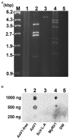
Figure 1
Figure 1
Detection of the 5' cap structures in the AaV1 dsRNA genome by RNA dot blot with the anti-m7G-Cap mAb. (A) Viral dsRNAs from purified virus particles. The dsRNAs were isolated from virus particles and then electrophoresed in 1% agarose gel with EtBr (0.5μg/ml) at 18V for 20h. Lane designation: M, 250ng of λ-EcoT14I-digested DNA marker; 1, AaV1-free; 2, AaV1 dsRNAs; 3, ScV-L-A dsRNA; 4, MyRV1 dsRNAs; 5, DW (distilled water, no template control). (B) RNA dot blot assay with the anti-m7G-Cap mAb. The dsRNA solutions (1,000, 500, and 250ng/μl) were dropped on the nylon membrane then probed with anti-m7G-Cap mAb. The AaV1 dsRNAs and the MyRV1 dsRNAs (positive control) showed positive signals, while the AaV1-free sample, the ScV-L-A dsRNA (negative control) and DW showed no signal.
Figure 2
Figure 2
Detection of 5' cap structures on each AaV1 dsRNA segment by RLM-RACE. (A) Electrophoresis of the separately excised AaV1 dsRNA segments in a 1% agarose gel with EtBr (0.5μg/ml) at 50V for 1h (Mupid-2plus, Takara Bio, Japan). (B) Results of the RLM-RACE analysis, confirming the presence of 5' cap structures on each AaV1 dsRNA segment. The separately excised dsRNAs were subjected to the RLM-RACE procedure (Supplementary Figure S3) and then electrophoresed in a 1% agarose gel with EtBr (0.5μg/ml) at 100V for 0.5h. Lane M, 100bp DNA ladder. The arrows indicate the amplified target bands. (C) Diagrams of the AaV1 dsRNA1, 2, 3, and 4 segments showing the primer pairs used for RLM-RACE and for amplification of each full-length dsRNA segment.
Figure 3
Figure 3
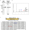
Characterization of the AaV1 major coat protein. (A) SDS-PAGE and western blot of the purified AaV1 particles. Purified viral proteins from strain EGS 35–193-1d were stained with CBB (left) or immunoblotted with antiserum against the AaV1 virus particles (right). The arrows indicate the viral structural protein. (B) Reverse-phase HPLC of the 82kDa major protein after digestion with Lysyl endopeptidase and trypsin. Peaks 2 and 3 were subjected to Edman degradation. (C) The deduced amino acid sequence of ORF3 (759 aa) written in one-letter code. The peptide sequences of peak 2 (blue) and peak 3 (red) were the same as the two regions in the predicted ORF3 peptide sequence.
Figure 4
Figure 4
Phenotypic changes in EGS 35–193 mycelia caused by deletions of AaV1 dsRNA2. (A) Four types of colony morphologies were exhibited by the EGS 35–193 derivatives EGS 35–193-1d (the original strain), EGS 35–193-0d, EGS 35–193-2d, and EGS 35-195-VF (AaV1-free), grown on YGA plates for 7days at 25°C. (B, C) Agarose gel electrophoresis of dsRNAs purified from mycelia (20mg) of the four EGS 35–193 derivatives, purified by the spin column method (B), and dsRNAs extracted from purified virus particles of the three EGS 35–193 AaV1-infected isolates (C). The dsRNAs were separated in 1.0% agarose gels with EtBr (0.5μg/ml) at 18V for 20h. Lane designation: M, 250ng of λ-EcoT14I-digested DNA marker; 1d, EGS 35–193-1d; 0d, EGS 35–193-0d; 2d, EGS 35–193-2d; VF, EGS 35-193-VF. (D) SDS-PAGE of purified virus particles from EGS 35–193-1d, −0d, and-2d. The purified viral proteins were separated in an 8% polyacrylamide gel at 120V for 2h and then stained with CBB. Lane M, prestained protein marker. (E) Western blot analysis of purified virus particles from EGS 35–193-1d, −0d, and-2d, with antiserum raised against the AaV1 virus particles from EGS 35–193-1d.
Figure 5
Figure 5
Analysis of the AaV1 dsRNA2-associated segments in virons purified from the three AaV1-infected isolates. (A-C) RT-PCR detection of the full-length dsRNA1–4 segments in EGS 35–193-1d (A), EGS 35–193-0d (B), and EGS 35–193-2d (C). RT-PCR was performed with the four primer pairs (Figure 2C; Supplementary Table S1) designed to amplify the full-length dsRNA1–4 segments. The RT-PCR products were separated in 1.0% agarose gels with EtBr (0.5μg/ml) at 50V for 1h. Lane designation: M, 250ng of λ-EcoT14I-digested DNA marker; 1, dsRNA1; 2, dsRNA2; 3, dsRNA3; 4, dsRNA4. (D) Position of the DIG DNA probe used to detect the 3' region of AaV1 dsRNA2 in the northern hybridization experiment. The DIG DNA probe was synthesized using the probe synthesis primer pair (Supplementary Table S1). (E) Agarose gel electrophoresis of dsRNA genomes extracted from the purified virus particles of the three AaV1-infected isolates. These AaV1 dsRNAs were separated in a 1.0% agarose gel with EtBr (0.5μg/ml) at 18V for 20h. Lane M, 250ng of λ-EcoT14I-digested DNA marker. (F) Northern hybridization to detect the AaV1 dsRNA2-associated segments. After agarose gel electrophoresis, the dsRNA genomes were denatured and blotted onto a nylon membrane, and probed with the DIG DNA probe. (G) Relative quantification of the three dsRNA2 segments, intact, del-1 and del-2, following northern hybridization (F). The total signal in each lane was normalized using the amount of dsRNA 1, following agarose gel electrophoresis (E). Quantification of individual bands was performed using Fiji/ImageJ.
Figure 6
Figure 6
Determination of the deleted regions in the dsRNA2 segments of AaV1. (A) Agarose gel electrophoresis of RT-PCR products amplified with the primer pair AaV1 dsRNA2-F and AaV1 dsRNA2-R, which is designed to amplify full-length AaV1 dsRNA2. The 1.0% agarose gel with EtBr (0.5μg/ml) was run at 18V for 20h. Lane M, 250ng of λ-EcoT14I-digested DNA marker. (B, C) Schematic diagrams showing the genome deletions of the AaV1 dsRNA2 derivatives. (B) Defective dsRNA2 del-1 has an in-frame deletion site (D1, 231bp) located at nt 1,275–1,505 of the intact AaV1 dsRNA2. (C) Defective dsRNA2 del-2 has two in-frame deletion sites, D1 and D2 (465bp), located at nt 113–577 of the intact AaV1 dsRNA2.

References

    1. Aoki N., Moriyama H., Kodama M., Arie T., Teraoka T., Fukuhara T. (2009). A novel mycovirus associated with four double-stranded RNAs affects host fungal growth in Alternaria alternata. Virus Res. 140, 179–187. doi: 10.1016/j.virusres.2008.12.003, PMID: - DOI - PubMed
    1. Arnesen T., Van Damme P., Polevoda B., Helsens K., Evjenth R., Colaert N., et al. . (2009). Proteomics analyses reveal the evolutionary conservation and divergence of N-terminal acetyltransferases from yeast and humans. Proc. Natl. Acad. Sci. U. S. A. 106, 8157–8162. doi: 10.1073/pnas.0901931106 - DOI - PMC - PubMed
    1. Cai G., Myers K., Fry W. E., Hillman B. I. (2019). Phytophthora infestans RNA virus 2, a novel RNA virus from Phytophthora infestans, does not belong to any known virus group. Arch. Virol. 164, 567–572. doi: 10.1007/s00705-018-4050-0, PMID: - DOI - PubMed
    1. Celik A., Baker R., He F., Jacobson A. (2017). High-resolution profiling of NMD targets in yeast reveals translational fidelity as a basis for substrate selection. RNA 23, 735–748. doi: 10.1261/rna.060541.116, PMID: - DOI - PMC - PubMed
    1. Desselberger U. (1996). Genome rearrangements of rotaviruses. Adv. Virus Res. 46, 69–95. doi: 10.1016/s0065-3527(08)60070-6, PMID: - DOI - PubMed