Review
. 2021 Oct 20:12:773352.
doi: 10.3389/fimmu.2021.773352. eCollection 2021.

Dermatomyositis With Anti-MDA5 Antibodies: Bioclinical Features, Pathogenesis and Emerging Therapies

Affiliations
Review

Dermatomyositis With Anti-MDA5 Antibodies: Bioclinical Features, Pathogenesis and Emerging Therapies

Anaïs Nombel et al. Front Immunol. .

Abstract

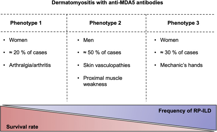
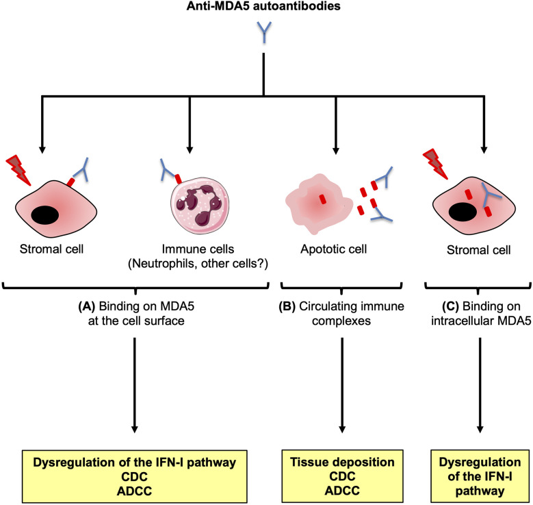
Anti-MDA5 dermatomyositis is a rare systemic autoimmune disease, historically described in Japanese patients with clinically amyopathic dermatomyositis and life-threatening rapidly progressive interstitial lung disease. Subsequently, the complete clinical spectrum of the disease was enriched by skin, articular and vascular manifestations. Depending on the predominance of these symptoms, three distinct clinical phenotypes with different prognosis are now defined. To date, the only known molecular component shared by the three entities are specific antibodies targeting MDA5, a cytosolic protein essential for antiviral host immune responses. Several biological tools have emerged to detect these antibodies, with drawbacks and limitations for each of them. However, the identification of this highly specific serological marker of the disease raises the question of its role in the pathogenesis. Although current knowledge on the pathogenic mechanisms that take place in the disease are still in their enfancy, several lines of evidence support a central role of interferon-mediated vasculopathy in the development of skin and lung lesions, as well as a possible pathogenic involvement of anti-MDA5 antibodies. Here, we review the clinical and biological evidences in favor of these hypothesis, and we discuss the contribution of emerging therapies that shed some light on the pathogenesis of the disease.

Keywords: COVID-19; MDA5; SARS-CoV-2; autoantibodies; autoantibody; dermatomyositis; idiopathic inflammatory myopathies; myositis.


Conflict of interest statement

The authors declare that the research was conducted in the absence of any commercial or financial relationships that could be construed as a potential conflict of interest.

Figures

Figure 1
Figure 1
The clinical phenotypes of anti-MDA5 dermatomyositis. Anti-MDA5 dermatomyositis can be divided into three clinical phenotypes with varying degrees of pulmonary damage, which is inversely correlated with the survival. RP-ILD, rapidly progressive interstitial lung disease.
Figure 2
Figure 2
Indirect immunofluorescence patterns of HEp-2 cells stained with anti–MDA5 positive sera. Stainings were performed on HEp-2 cells (Kallestad, Biorad) with sera from patients with anti-MDA5 Abs dermatomyositis. Top left: typical fine granular cytoplasmic staining in rare clustered cells. Top right: granular cytoplasmic pattern in all cells (which can mask the typical cytoplasmic pattern). Bottom left: nuclear speckled pattern, associated with the typical cytoplasmic pattern. Bottom right: isolated nuclear speckled pattern. Note that absence of fluorescence can also be observed with some sera.
Figure 3
Figure 3
Possible genetic and environmental factors involved in the anti-MDA5 dermatomyositis. Viral double stranded RNA (dsRNA) activates MDA5 in infected cells, leading to type I interferon (IFN-I) production and increased levels of MDA5. Altered WDFY4 impairs antigen cross-presentation by CD1c+ dendritic cells (DCs), favoring an inefficient elimination of infected cells and further activation of MDA5. In parallel, altered WDFY4 also enhances MDA5-mediated nuclear factor-kappa B (NF-κB) pathway leading to the apoptosis of infected cells, and the release of MDA5. Local dysfunctional mitochondrial polynucleotide phosphorylase (PNPase) could lead to intracellular accumulation of endogenous dsRNA, fueling uncontrolled activation and expression of MDA5. Abnormal accumulation of MDA5 may favor a loss of tolerance to MDA5 in an individual with proper genetic background, leading to anti-MDA5 antibodies (Abs).
Figure 4
Figure 4
Endothelial dysfunctions and immune alterations in anti-MDA5 dermatomyositis. In the skin, CXCL10, a keratinocyte and endothelial-derived chemokine, induces the recruitment of CD8+CXCR3+ T cells, potentially autoreactive and leading to keratinocytes death. Endothelin released by injured endothelial cells, is a strong vasoconstrictor which can induce local ischemia responsible for cutaneous ulcers. In the lung, CX3CL1 can be produced by vascular pulmonary endothelial cells following IFN-I exposure. CX3CL1 recruits CX3CR1+ alternative alveolar macrophages (M2). Alveolar M2 macrophages, as well as airway epithelial cells, release stromal cell-derived factor 1 (SDF-1) which induces the accumulation of intrapulmonary CD4+CXCR4+ T cells. CD4+CXCR4+ T cells produce profibrotic agents (transformation growth factor-β (TGF-β), α-smooth muscle actin (α-SMA) and collagen I), as well as IL-21, which promotes the differentiation of profibrotic CD8+ T cells. CD8+ T cells secrete IL-13, which stimulates macrophages to produce profibrotic factors.
Figure 5
Figure 5
Potential contribution of the anti-MDA5 antibodies to the pathogenesis. Anti-MDA5 antibodies (Abs) may contribute to the pathogenesis in several ways. (A) In specific conditions, MDA5 may translocate at the surface of critical stromal cells, or immune cells such as neutrophils. Interaction between the autoAb and the ectopic antigenic target could trigger chronic activation of the type I interferon (IFN-I) signaling pathway, as well as immune mediated cytotoxicity through complement activation (CDC) and/or Ab-dependent cytotoxicity (ADCC). (B) Anti-MDA5 Abs could also bind to MDA5 released from apoptotic cells, to form immune complexes that could contribute to immune-mediated damage. (C) A cell exposed to a stress (infection, genetic background) overexpresses intracytoplasmic MDA5. Anti-MDA5 Abs might penetrate the cell to bind to MDA5, altering several functional pathways.

References

    1. Mariampillai K, Granger B, Amelin D, Guiguet M, Hachulla E, Maurier F, et al. . Development of a New Classification System for Idiopathic Inflammatory Myopathies Based on Clinical Manifestations and Myositis-Specific Autoantibodies. JAMA Neurol (2018) 75:1528–37. doi: 10.1001/jamaneurol.2018.2598 - DOI - PMC - PubMed
    1. Allenbach Y, Uzunhan Y, Toquet S, Leroux G, Gallay L, Marquet A, et al. . Different Phenotypes in Dermatomyositis Associated With Anti-MDA5 Antibody: Study of 121 Cases. Neurology (2020) 95:e70–8. doi: 10.1212/WNL.0000000000009727 - DOI - PMC - PubMed
    1. Betteridge Z, Tansley S, Shaddick G, Chinoy H, Cooper RG, New RP, et al. . Frequency, Mutual Exclusivity and Clinical Associations of Myositis Autoantibodies in a Combined European Cohort of Idiopathic Inflammatory Myopathy Patients. J Autoimmun (2019) 101:48–55. doi: 10.1016/j.jaut.2019.04.001 - DOI - PMC - PubMed
    1. Sato S, Hirakata M, Kuwana M, Suwa A, Inada S, Mimori T, et al. . Autoantibodies to a 140-Kd Polypeptide, CADM-140, in Japanese Patients With Clinically Amyopathic Dermatomyositis. Arthritis Rheum (2005) 52:1571–6. doi: 10.1002/art.21023 - DOI - PubMed
    1. Fujikawa K, Kawakami A, Kaji K, Fujimoto M, Kawashiri S, Iwamoto N, et al. . Association of Distinct Clinical Subsets With Myositis-Specific Autoantibodies Towards Anti-155/140-kDa Polypeptides, Anti-140-kDa Polypeptides, and Anti-Aminoacyl tRNA Synthetases in Japanese Patients With Dermatomyositis: A Single-Centre, Cross-Sectional Study. Scand J Rheumatol (2009) 38:263–7. doi: 10.1080/03009740802687455 - DOI - PubMed

Publication types