. 2022 Jan;9(1):e2101553.
doi: 10.1002/advs.202101553. Epub 2021 Nov 7.

Citrate Promotes Excessive Lipid Biosynthesis and Senescence in Tumor Cells for Tumor Therapy

Affiliations

Citrate Promotes Excessive Lipid Biosynthesis and Senescence in Tumor Cells for Tumor Therapy

Yangjing Zhao et al. Adv Sci (Weinh). 2022 Jan.

Abstract

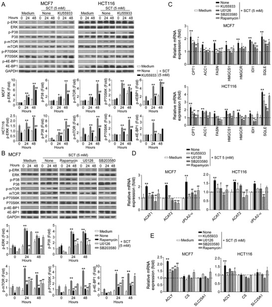
Metabolic disorder is one of the hallmarks of cancers, and reprogramming of metabolism is becoming a novel strategy for cancer treatment. Citrate is a key metabolite and critical metabolic regulator linking glycolysis and lipid metabolism in cellular energy homeostasis. Here it is reported that citrate treatment (both sodium citrate and citric acid) significantly suppresses tumor cell proliferation and growth in various tumor types. Mechanistically, citrate promotes excessive lipid biosynthesis and induces disruption of lipid metabolism in tumor cells, resulting in tumor cell senescence and growth inhibition. Furthermore, ATM-associated DNA damage response cooperates with MAPK and mTOR signaling pathways to control citrate-induced tumor cell growth arrest and senescence. In vivo studies further demonstrate that citrate administration dramatically inhibits tumor growth and progression in a colon cancer xenograft model. Importantly, citrate administration combined with the conventional chemotherapy drugs exhibits synergistic antitumor effects in vivo in the colon cancer models. These results clearly indicate that citrate can reprogram lipid metabolism and cell fate in cancer cells, and targeting citrate can be a promising therapeutic strategy for tumor treatment.

Keywords: DNA damage response; MAPK; apoptosis; cellular senescence; chemotherapy; citrate; lipid metabolism; mTOR.


Conflict of interest statement

The authors declare no conflict of interest.

Figures

Figure 1
Figure 1
Increased ACLY expression in various tumor tissues is associated with poor clinical outcomes in cancer patients. A) ACLY mRNA expression was upregulated in tumor tissues compared with normal samples detected by RNA sequencing in eight tumor types. The sequencing data were obtained from the TCGA database. Black dots represent normal samples and red dots represent tumor samples. Data shown are mean ± SD. **p<0.01, between normal and tumor samples in each tumor type. B) OS and RFS analyses were performed in patients with low or high ACLY mRNA expression in six tumor types. The sequencing data were obtained from the TCGA database. The Kaplan–Meier plots were analyzed by the log‐rank test. C) High mRNA expression of ACLY in cancer cell lines compared with normal cell lines (WI‐38 and MCF10A). Total RNA was isolated from cells and gene expression was analyzed by real‐time qPCR. ACLY expression levels were normalized to β‐actin expression level and adjusted to the levels in WI‐38 cells (served as 1). **p<0.01, compared with WI‐38 cells. D) Inhibition of ACLY induced increases of intracellular CA in both human normal cell lines (WI‐38 and MCF10A) and multiple cancer cell lines. Citrate levels in indicated cells were detected by CA content assay kit after being treated with or without ACLY inhibitor SB204990 (10 µm) for 48 h. **p<0.01, compared with WI‐38 cells. # p<0.05 and ## p<0.01, compared with the respective medium‐only group. Unpaired Student's t‐test was performed in (A). Log‐rank test was used to determine the statistical significance in (B). One‐way analysis of variance (ANOVA) was performed in (C) and (D).
Figure 2
Figure 2
SCT inhibits tumor cell proliferation and growth. A) Multiple cancer cell lines were treated with increasing doses of SCT for 24 h and cell proliferation was measured by the MTT assay. The normal HFF, WI‐38, and MCF10A cells served as controls. The values of cell proliferation are shown as mean ± SD of six repeated wells. IC50 value for each cell type is a representative of three independent experiments. B) MCF7, HCT116, HT29, and A549 tumor cells and normal HFF, WI‐38, and MCF10A cells were seeded at optimized starting numbers in 24‐well plates in the presence of indicated concentrations of SCT. The cell growth was evaluated at different time points using cell number counting. Data shown are mean ± SD from three independent experiments. **p<0.01, compared with the medium‐only group. C,D) Low doses of SCT did not induce tumor cell apoptosis. Different types of tumor cells were treated with the indicated concentrations of SCT for 48 h. Normal HFF, WI‐38, and MCF10A cells were included as a control. Apoptotic cells were analyzed using the flow cytometric analysis after Annexin V and 7‐AAD double staining. Data shown (in D) are mean ± SD from three experiments with similar results. *p<0.05 and **p<0.01, compared with the medium‐only group. E) Only high dose of SCT induced apoptotic protein expression in tumor cells. HCT116 and H1299 cells were treated with/without the indicated concentrations of SCT for 24 h. Protein expressions of cleaved caspase‐3, caspase‐3, Bax, Bcl‐2, and Mcl‐1 were determined using the western blot analyses. ANOVA was performed in (B) and (D).
Figure 3
Figure 3
SCT induces DDRs and senescence in tumor cells. A) Extracellular SCT treatment increased senescent cell populations in tumor cells but not in normal cells. MCF7, HCT116, and WI‐38 cells were treated with the indicated concentrations of SCT for 24 h and then stained for SA‐β‐Gal. SA‐β‐Gal+ cells were shown with dark blue granules as indicated by the arrows. Data shown in histograms are mean ± SD from three independent experiments. *p<0.05 and **p<0.01, compared with the medium‐only group. Scale bar: 30 µm. B,C) Phosphorylated activations of key DNA damage molecules ATM, H2AX, 53BP1, and CHK2 in MCF7 and HCT116 tumor cells were detected by the flow cytometry analysis (in B) and the immunofluorescence staining (in C) after culture with indicated concentrations of SCT for 24 h. Scale bar: 100 µm. D,E) Inhibition of ATM signaling reversed citrate‐induced suppression on tumor cell proliferation and growth. MCF7 and HCT116 cells were pretreated with ATM inhibitor KU55933 (5 µm) for 24 h and then cultured with SCT (5 mm) for 48 h. Cell proliferation and growth were determined with the MTT assay (in D) and cell numbers counting (in E), respectively. Proliferation of tumor cells with medium only reversed as 100% (in C). Data shown are mean ± SD from three independent experiments. **p<0.01, compared with the medium‐only group. ## p<0.01, compared with citrate treatment only group. (F) Blockage of ATM signaling prevented citrate‐induced tumor cell senescence. Cell treatment and procedure were identical to (D) and (E). Senescent cell populations were determined using the SA‐β‐Gal staining. Data shown in histograms are mean ± SD from three independent experiments. **p<0.01, compared with the medium‐only group. ## p<0.01, compared with the citrate treatment group. Scale bar: 30 µm. ANOVA was performed in (A), (D), (E), and (F).
Figure 4
Figure 4
SCT‐induced excessive lipid biosynthesis is responsible for cell senescence and suppression in tumor cells. A) Increased gene expression levels of key enzymes in cholesterol synthesis (HMGCR, HMGCS1, SQLE, and IDI1), as well as fatty acid oxidation (CPT‐1) and synthesis (ACC1 and FASN) in tumor cells were induced with SCT treatment for 24 h. Total RNA was isolated from the treated tumor cells and gene expression was analyzed by real‐time qPCR. Expression levels of each gene were normalized to β‐actin expression level and adjusted to the levels in tumor cells treated with medium only (served as 1). Data shown are mean ± SD from three independent experiments with similar results. *p<0.05 and **p<0.01, compared with the medium‐only group. B) Accumulated LDs in tumor cells induced by SCT treatment. MCF7 and HCT116 cells were cultured in the presence of indicated concentrations of SCT for 24 or 48 h. The treated tumor cells were performed Oil Red O staining. The Oil Red O + tumor cells were identified with red granules as indicated by the arrows. Data shown in the right panels are means ± SD from three independent experiments. **p<0.01, compared with the medium‐only group. Scale bar: 30 µm. C) Schematic diagram of the lipid biosynthesis pathways. The key enzymes are shown in blue color and the specific pharmacological inhibitors used in this study are shown in red color. D) SCT treatment upregulated gene expression of key enzymes (ACAT1, ACAT2, and cPLA2α) involved in LD formation, but not hydrolase LIPA in tumor cells. MCF7 and HCT116 cells were treated with SCT (5 mm) for 24 h and mRNA expression levels of each gene were determined by the real‐time qPCR. The expression levels were normalized to β‐actin expression and adjusted to the levels in tumor cells with medium only. Data are mean ± SD from three independent experiments with similar results. *p<0.05 and **p<0.01, compared with the tumor cells in the respective medium only group. E) Blockage of the lipid synthesis reversed SCT‐induced LD accumulation in tumor cells. MCF7 and HCT116 cells were pretreated with the pharmaceutical inhibitors for lipid synthesis for 24 h, including C75 (5 µm), simvastatin (1 µm), 25‐HC (0.25 µg mL−1), or avasimible (1 µm), respectively. Tumor cells were then cultured in the presence of SCT (5 mm) for an additional 24 h and stained for Oil Red O. Data shown in the right panels are mean ± SD from three independent experiments with similar results. *p<0.05 and **p<0.01, compared with the medium‐only group. ## p<0.01, compared with the citrate treatment only group. Scale bar: 30 µm. F,G) Inhibition of the lipid synthesis blocked citrate‐induced suppression on tumor cell proliferation and growth. Cell treatment and procedure were identical to (E). Cell proliferation and growth were determined with the MTT assay (in F) and cell numbers counting (in G), respectively. Proliferation of tumor cells with medium only served as 100% (in F). Data shown are mean ± SD from three independent experiments. **p<0.01, compared with the medium‐only group. # p<0.05 and ## p<0.01, compared with the citrate treatment only group. H) Blockage of the lipid synthesis prevented citrate‐induced tumor cell senescence. Cell treatment and procedure were identical to (E). Senescent cell populations were determined using the SA‐β‐Gal staining. Data shown in histograms are mean ± SD from three independent experiments. **p<0.01, compared with the medium‐only group. ## p<0.01, compared with the citrate treatment only group. Scale bar: 30 µm. ANOVA was performed in (A–H).
Figure 5
Figure 5
ERK and p38 MAPK signaling pathways control the molecular process of citrate‐induced tumor cell senescence. A) SCT treatment significantly induced phosphorylation of ERK1/2 and P38 in tumor cells. MCF7 and HCT116 cells were cultured for indicated time points with or without SCT (1 or 5 mm), and cell lysates were prepared for western blot analyses. Phosphorylated ERK1/2 and P38 protein levels shown in the histograms were quantitatively analyzed and compared against the GAPDH expression levels with a densitometer. Results shown are mean ± SD from three independent experiments. *p<0.05 and **p<0.01, compared with the medium‐only group. B,C) Pretreatment of tumor cells with ERK1/2 and P38 inhibitors U0126 and SB203580 significantly prevented citrate‐induced suppression of tumor cell proliferation and growth. MCF7 and HCT116 cells were pretreated with or without U0126 (4 µm) or SB203580 (5 µm) for 24 h and then further cultured in the presence of SCT or CA for 48 h. Cell proliferation and growth were determined using the MTT (in B) and cell numbers counting (in C) assays, respectively. Data shown are mean ± SD from three independent experiments. **p<0.01, compared with the medium‐only group. ## p<0.01, compared with the citrate treatment only group. D) Blockage of the ERK1/2 and P38 signaling prevented citrate‐induced tumor cell senescence. MCF7 and HCT116 cells were pretreated with or without U0126 (4 µm) or SB203580 (5 µm) for 24 h and further cultured in the presence of SCT or CA for 24 h and then stained for SA‐β‐Gal. Data shown in the right panels are mean ± SD from three independent experiments. **p<0.01, compared with the medium‐only group. ## p<0.01, compared with the citrate treatment only group. Scale bar: 30 µm. E) Blockage of the ERK1/2 and P38 prevented citrate‐induced LD accumulation in tumor cells. Cell treatment and procedure were identical to (D). Oil Red O staining was performed to detect LD accumulation. Data shown in the histograms are mean ± SD from three independent experiments. **p<0.01, compared with the medium‐only group. ## p<0.01, compared with the citrate treatment only group. Scale bar: 30 µm. ANOVA was performed in (A–E).
Figure 6
Figure 6
mTOR signaling involves tumor cell senescence induced by citrate treatment. A) Phosphorylated activation of mTOR signaling pathway in tumor cells was induced by SCT treatment. Treatment and procedure of MCF7 and HCT116 cells were identical to (Figure 5A). Phosphorylated mTOR, p70, and 4E‐BP1 protein levels shown in the right histograms were quantitatively analyzed and compared against the GAPDH expression levels with a densitometer. Results shown are mean ± SD from three independent experiments. *p<0.05 and **p<0.01, compared with the medium‐only group. B,C) Inhibition of mTOR signaling by pharmacological inhibitor rapamycin markedly reversed citrate‐induced inhibition of tumor cell proliferation and growth. MCF7 and HCT116 cells were pretreated with rapamycin (50 nm) for 24 h, and then cultured in the presence of SCT (5 mm) or CA (4 mm) for an additional 48 h. Cell proliferation and growth were determined by the MTT assay (in B) and cell numbers counting (in C), respectively. Data shown are mean ± SD from three independent experiments. **p<0.01, compared with the medium‐only group. ## p<0.01, compared with the citrate treatment only group. D) Pretreatment of tumor cells with rapamycin significantly prevented citrate‐induced tumor cell senescence. MCF7 and HCT116 cells were pretreated with or without rapamycin (50 nm) for 24 h and then further cultured in the presence of SCT or CA for 24 h. Senescent cell populations were determined using the SA‐β‐Gal staining. Data shown in the histograms are mean ± SD from three independent experiments. **p<0.01, compared with the medium‐only group. # p<0.05 and ## p<0.01, compared with the citrate treatment only group. Scale bar: 30 µm. E) Blockage of mTOR activation markedly prevented citrate‐induced LD accumulation in tumor cells. Cell treatment and procedure were identical to (D). Oil Red O staining was performed to detect LD accumulation. Data shown in the histograms are mean ± SD from three independent experiments. **p<0.01, compared with the medium‐only group. ## p<0.01, compared with the citrate treatment only group. Scale bar: 30 µm. ANOVA was performed in (A–E).
Figure 7
Figure 7
The causative regulations between DDR, MAPK, and mTOR signaling pathways for citrated‐induced excessive lipid metabolism and senescence in tumor cells. A) Treatment with ATM inhibitor KU55933 significantly prevented citrate‐induced ERK1/2, P38, mTOR, p70, and 4E‐BP1 phosphorylation in tumor cells using western blot analyses. MCF7 and HCT116 cells were pretreated with or without KU55933 (5 µm) for 24 h, and then cultured in the presence of SCT (5 mm) for 24 or 48 h. Protein levels of phosphorylated ERK1/2, P38, mTOR, p70, and 4EBP1 were quantitatively analyzed and compared against the GAPDH expression levels with a densitometer. Results shown in the histograms are mean ± SD from three independent experiments. *p<0.05 and **p<0.01, compared with the medium‐only group. # p<0.05 and ## p<0.01, compared with the citrate treatment only group. B) Inhibition of ERK1/2 or P38 signaling pathways by specific pharmacological inhibitors markedly blocked citrate‐induced mTOR signaling activation in tumor cells. MCF7 and HCT116 cells were pretreated with inhibitors rapamycin (50 nm), U0126 (4 µm), or SB203580 (5 µm) for 24 h, and then treated with SCT (5 mm) for different time points. Phosphorylation of ERK1/2, P38 mTOR, p70, and 4EBP1 was determined by western blot analyses. Results shown in the histograms are mean ± SD from three independent experiments. **p<0.01, compared with the medium‐only group. # p<0.05 and ## p<0.01, compared with the citrate treatment only group. C) Inhibition of MAPK or mTOR signaling by specific inhibitors significantly suppressed the increased gene expression levels of key enzymes in cholesterol and fatty acid synthesis (HMGCR, HMGCS1, SQLE, and IDI1, CPT‐1, ACC1, and FASN) in tumor cells induced by SCT treatment. Cell treatment and procedure were identical to (A) and (B). Gene expression in treated tumor cells was analyzed by real‐time qPCR. Expression levels of each gene were normalized to β‐actin expression level and adjusted to the levels in tumor cells treated with medium only (served as 1). Data shown are mean ± SD from three independent experiments with similar results. *p<0.05 and **p<0.01, compared with the medium‐only group. # p<0.05 and ## p<0.01, compared with the citrate treatment only group. D,E) Blockage of the MAPK or mTOR signaling reversed the SCT‐induced upregulation of key enzymes (ACAT1, ACAT2, and cPLA2α) involved in LD formation (in D), and enzymes in citrate hydrolysis (in E) in tumor cells. Cell treatment, procedure, and data analysis were identical to (C). Data shown are mean ± SD from three independent experiments with similar results. *p<0.05 and **p<0.01, compared with the medium only group. # p<0.05 and ## p<0.01, compared with the citrate treatment only group. ANOVA was performed in (A–E).
Figure 8
Figure 8
SCT inhibits tumor growth and progression in vivo in a colon cancer xenograft model. A,B) SCT treatment dramatically inhibited tumor growth in Rag1−/−‐immunodeficient mice. HCT116 cells (4.2 × 10⁶/mouse) were subcutaneously injected into Rag1−/− mice. After 6 days of tumor injection, solvent control or SCT (15 mg kg−1 or 30 mg kg−1 body weight) were given by intraperitoneal injection at every other day for 16 days. Tumor volumes were measured and presented as mean ± SD (in A)(n = 9 mice/group). Representative images of the xenograft tumors obtained from the indicated groups at the endpoint of the experiments (day 22) are shown (in B). In addition, results shown are mean ± SD of the tumor weights from the indicated groups at the endpoint of the experiments (n = 9 mice/group). **p<0.01, compared with the solvent control injection group. C) SCT treatment significantly decreased Ki‐67+ cell populations in the tumor tissues using an immunofluorescence assay. Left panels are representative images of Ki‐67 expression in tumor tissues from different groups. Right panel is the summary of mean ± SD of Ki‐67+ cell fractions per high microscope field (× 400) in the tumor tissues from 9 mice of each group. **p<0.01, compared with the PBS control treatment mice. Scale bar: 30 µm. D) SCT treatment did not induce cleaved caspase‐3+ cell populations in tumor tissues using the immunofluorescence assay. Left panels are representative images of cleaved caspase‐3 expression in frozen tumor tissues from different groups. Right panel is the summary of mean ± SD of cleaved caspase‐3+ cell fractions per high microscope field (× 400) in the tumor tissues from 9 mice of each group. Scale bar: 30 µm. E) Large amounts of senescent tumor cells were observed in tumor tissues in SCT‐treated mice. SA‐β‐Gal expression was determined in the tumor frozen tissues from different groups at the endpoint of the experiment. Data shown in the histograms are mean ± SD of SA‐β‐Gal+ cell numbers per high microscope field (× 400) in the tumor tissues from 9 mice of each group. **p<0.01, compared with the control group. Scale bar: 30 µm. F) SCT treatment dramatically increased LD accumulation in tumor tissues from tumor‐bearing mice. Data shown in the lower histograms are mean ± SD of Oil Red O+ cell numbers per high microscope field (× 400) in the tumor tissues from 9 mice of each group. **p<0.01, compared with the control group. Scale bar: 30 µm. G) SCT administration increased gene expression of key enzymes involved in cholesterol and fatty acid synthesis (HMGCR, HMGCS1, SQLE, and IDI1, CPT‐1, ACC1, and FASN) and LD formation (ACAT1, ACAT2, and cPLA2α) in tumor tissues using the real‐time qPCR analyses. Expression levels of each gene were normalized to β‐actin expression levels and adjusted to the levels in the control group (served as 1). Data shown are mean ± SD from the tumor tissues from 9 mice of each group. *p<0.05 and **p<0.01, compared with the control group. H) SCT administration induced the phosphorylation of ERK1/2 and P38 in tumor tissues using western blot analyses. Phosphorylated ERK1/2 and P38 protein levels shown in the lower scatter diagrams were quantitatively analyzed and compared against the GAPDH expression levels with a densitometer. Every single dot represents one sample from an individual mouse. Results shown in the scatter diagrams are mean ± SD from three independent experiments. *p<0.05 and **p<0.01, compared with the control group. ANOVA was performed in (A–H).
Figure 9
Figure 9
SCT administration synergistically enhances the antitumor efficacy of traditional chemotherapy. A,B) SCT treatment combined with chemotherapeutic agents showed synergistically enhanced inhibitory activity on tumor cell proliferation and growth. MCF7 and HCT116 cells were treated with SCT (5 mm) in combination with or without 5‐FU (300 µm), paclitaxel (5 µm), or oxaliplatin (50 µm). Cell proliferation and growth were determined with the MTT assay (in A) and cell numbers counting (in B), respectively. Proliferation of tumor cells with medium only was set as 100% (in A). Data shown are mean ± SD from three independent experiments. **p<0.01, compared with the medium‐only group. ## p<0.01, compared with the citrate treatment only group. C–F) Combination with SCT treatment improved the anti‐tumor efficacy of chemotherapeutic agents in vivo in the HCT116 tumor models. HCT116 cells (4.2 × 10⁶/mouse) were subcutaneously injected into Rag1−/− mice. Tumor‐bearing mice received solvent control, SCT (15 mg kg−1 body weight), 5‐FU (20 mg kg−1 body weight), or oxaliplatin (10 mg kg−1 body weight) by intraperitoneal injection. Tumor volumes shown in (C) and (E) were measured and presented as mean ± SD (n = 4–5 mice/group). Representative images of the xenograft tumors obtained from the indicated groups at the endpoint of the experiments (day 22) are shown in (D) and (F). In addition, histogram results shown in (D) and (F) are mean ± SD of the tumor weights from the indicated groups at the endpoint of the experiments. Each dot represents one tumor from one mouse. **p<0.01, compared with the solvent control group. G) SCT treatment combined with 5‐FU or oxaliplatin had more inhibitory activity on tumor cell proliferation than that of SCT treatment only group. Tumor frozen sections were subjected to immunofluorescence staining for Ki‐67 expression. Upper panels are representative images of Ki‐67 expression in tumor tissues from different groups. Results shown in the lower histogram are mean ± SD of Ki‐67+ cell fractions per high microscope field (× 400) in the tumor tissues from each group (n = 4–5 mice/group). **p<0.01, compared with the control group. ## p<0.01, compared with the citrate treatment only group. Scale bar: 30 µm. H) SCT treatment combined with 5‐FU or oxaliplatin slightly increased apoptotic cells in tumor tissues. Cleaved caspase‐3 expression in tumor tissues was determined using the immunofluorescence assay. Left panels are representative images of cleaved caspase‐3 expression in frozen tumor tissues from different groups. Right panel is the summary of mean ± SD of cleaved caspase‐3+ cell fractions per high microscope field (× 400) in the tumor tissues from 4 mice of each group. # p<0.05, compared with the citrate‐treated only group. Scale bar: 30 µm. I,J) SCT treatment combined with 5‐FU or oxaliplatin promoted senescence induction (in I) and LD accumulation (in J) in tumor tissues than that of SCT treatment only group. SA‐β‐Gal and Oil Red O staining were performed in the tumor frozen tissues from different groups, respectively. Data shown in the histogram are mean ± SD of SA‐β‐Gal+/Oil Red O+ cell numbers per high microscope field (× 400) in the tumor tissues from 4 mice of each group. *p<0.05 and **p<0.01, compared with the control group. ## p<0.01, compared with the citrate‐treated only group. Scale bar: 30 µm. ANOVA was performed in (A–J).

References

    1. Ward P. S., Thompson C. B., Cancer Cell 2012, 21, 297. - PMC - PubMed
    1. Hay N., Nat. Rev. Cancer 2016, 16, 635. - PMC - PubMed
    1. Beloribi‐Djefaflia S., Vasseur S., Guillaumond F., Oncogenesis 2016, 5, e189. - PMC - PubMed
    1. Martinez‐Outschoorn U. E., Peiris‐Pages M., Pestell R. G., Sotgia F., Lisanti M. P., Nat. Rev. Clin. Oncol. 2017, 14, 11. - PubMed
    1. Zhao Y., Liu H., Liu Z., Ding Y., Ledoux S. P., Wilson G. L., Voellmy R., Lin Y., Lin W., Nahta R., Liu B., Fodstad O., Chen J., Wu Y., Price J. E., Tan M., Cancer Res. 2011, 71, 4585. - PMC - PubMed

Publication types

LinkOut - more resources